
















©The Author(s) 2025.
World J Clin Cases. Oct 26, 2025; 13(30): 109977
Published online Oct 26, 2025. doi: 10.12998/wjcc.v13.i30.109977
Published online Oct 26, 2025. doi: 10.12998/wjcc.v13.i30.109977
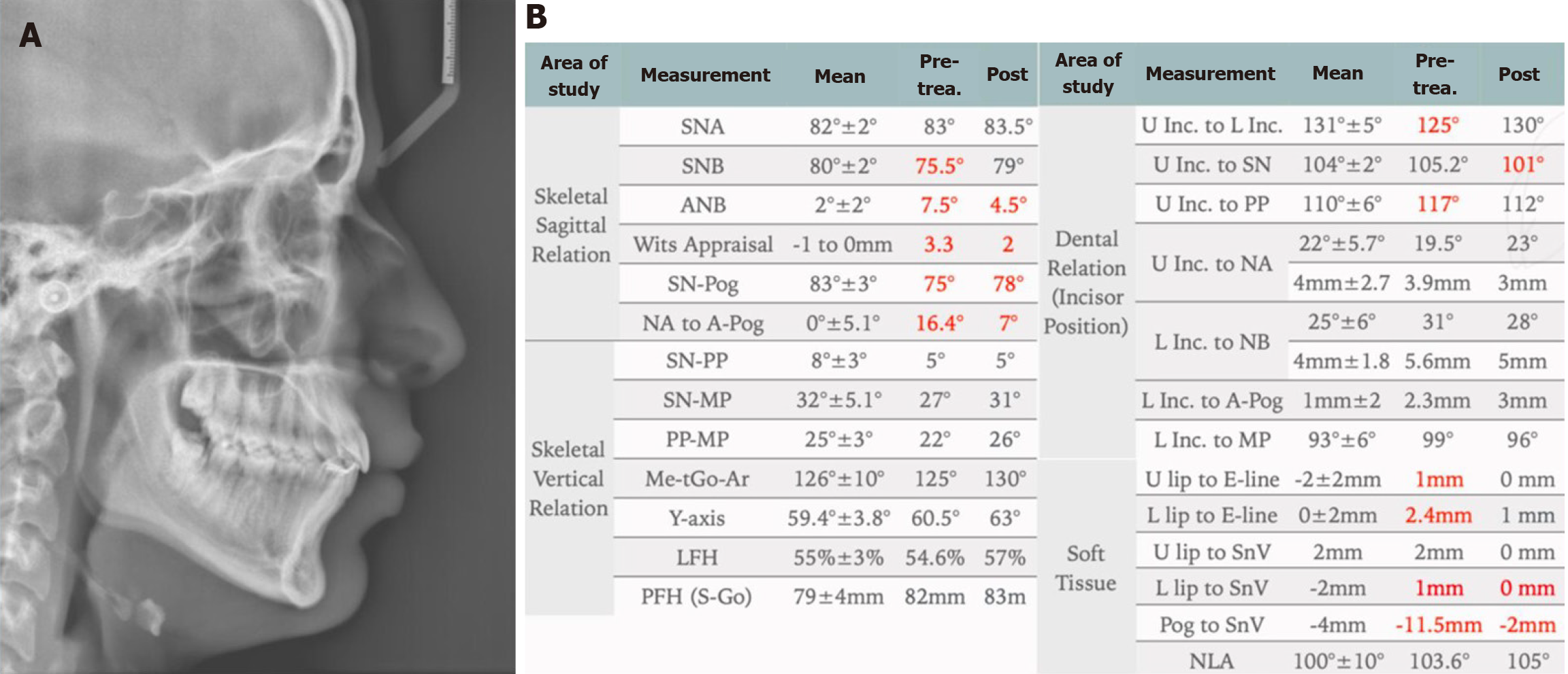
Figure 10 End of orthodontic treatment lateral cephalometric radiograph.
A: The lateral cephalometric radiograph shows significant improvements in skeletal and soft tissue positions, as well as in tooth positions; B: Cephalometric measurements for pre- and post-orthodontic treatment confirm that the overall improvement in skeletal and soft tissue measurements is maintained, as well as an improvement in the inclination of the upper and lower incisors.
- Citation: Alomair D, Halawani MR, Alsubaye S, Almajed AA. Treatment of class II malocclusion and ectopic maxillary canines: A case report. World J Clin Cases 2025; 13(30): 109977
- URL: https://www.wjgnet.com/2307-8960/full/v13/i30/109977.htm
- DOI: https://dx.doi.org/10.12998/wjcc.v13.i30.109977