
















©The Author(s) 2025.
World J Clin Cases. Oct 26, 2025; 13(30): 108828
Published online Oct 26, 2025. doi: 10.12998/wjcc.v13.i30.108828
Published online Oct 26, 2025. doi: 10.12998/wjcc.v13.i30.108828
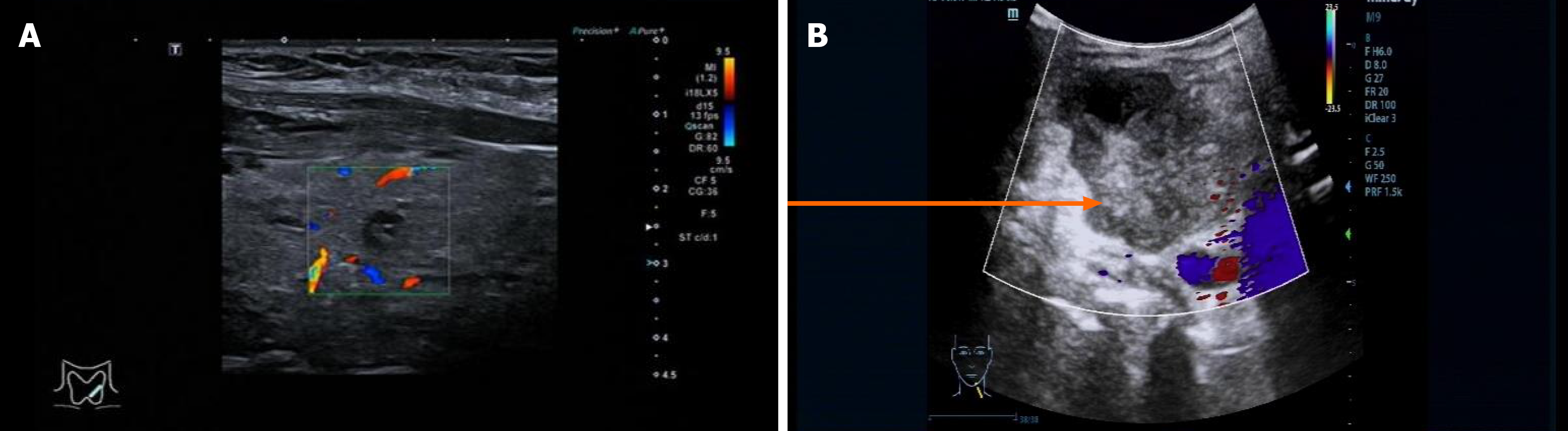
Figure 1 Thyroid ultrasound.
A: Right thyroid, hypoechoic nodule (TI-RADS 4b); B: Left lateral neck mass. A mixed echogenic mass measuring approximately 6.4 cm × 4.5 cm × 5.3 cm, with color Doppler flow imaging showing clear borders and regular shape.
- Citation: Li YL, Xia XL, Zhang SY, Tang L, Liu LN, Liu LS. Contralateral metastatic papillary thyroid carcinoma and complicated by primary hyperaldosteronism: A case report. World J Clin Cases 2025; 13(30): 108828
- URL: https://www.wjgnet.com/2307-8960/full/v13/i30/108828.htm
- DOI: https://dx.doi.org/10.12998/wjcc.v13.i30.108828