
















©The Author(s) 2025.
World J Clin Cases. Oct 26, 2025; 13(30): 108566
Published online Oct 26, 2025. doi: 10.12998/wjcc.v13.i30.108566
Published online Oct 26, 2025. doi: 10.12998/wjcc.v13.i30.108566
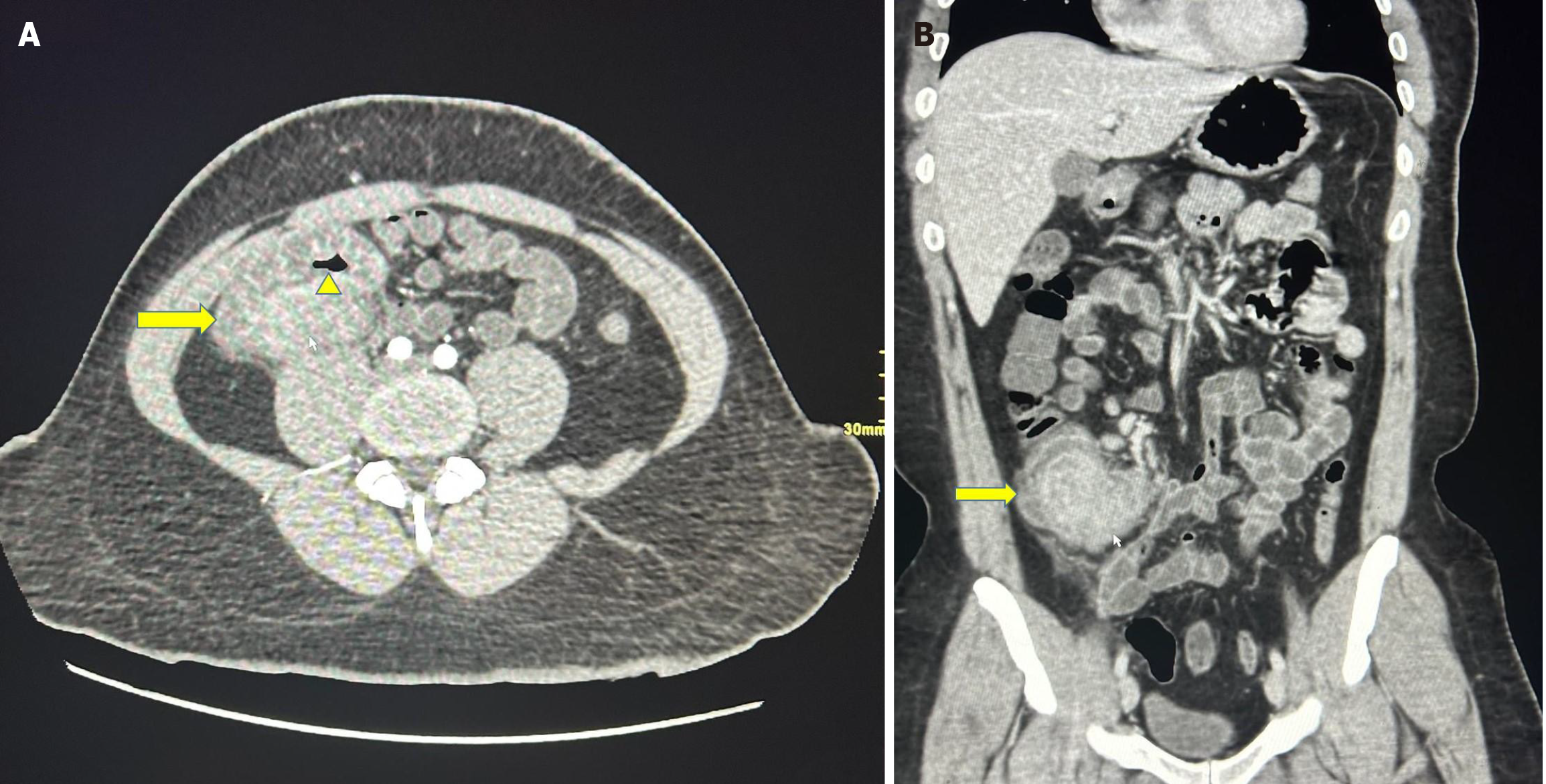
Figure 1 Computed tomography scan.
A: The axial section of the computed tomography (CT) scan shows a mass lesion involving the ileocecal region (yellow arrow) without complete obstruction of the intestinal lumen (yellow arrowhead); B: The coronal section of the CT scan shows a mass lesion involving the ileocecal region (yellow arrow).
- Citation: Alsulaimi MS, Memon MY, Alyabary IM, Almotairi E, Sulaiman T, Shamsan AN, Mubarak M. Ileocecal basidiobolomycosis mimicking malignancy successfully treated without surgery: A case report. World J Clin Cases 2025; 13(30): 108566
- URL: https://www.wjgnet.com/2307-8960/full/v13/i30/108566.htm
- DOI: https://dx.doi.org/10.12998/wjcc.v13.i30.108566