Copyright
©The Author(s) 2025.
World J Clin Cases. Sep 26, 2025; 13(27): 109243
Published online Sep 26, 2025. doi: 10.12998/wjcc.v13.i27.109243
Published online Sep 26, 2025. doi: 10.12998/wjcc.v13.i27.109243
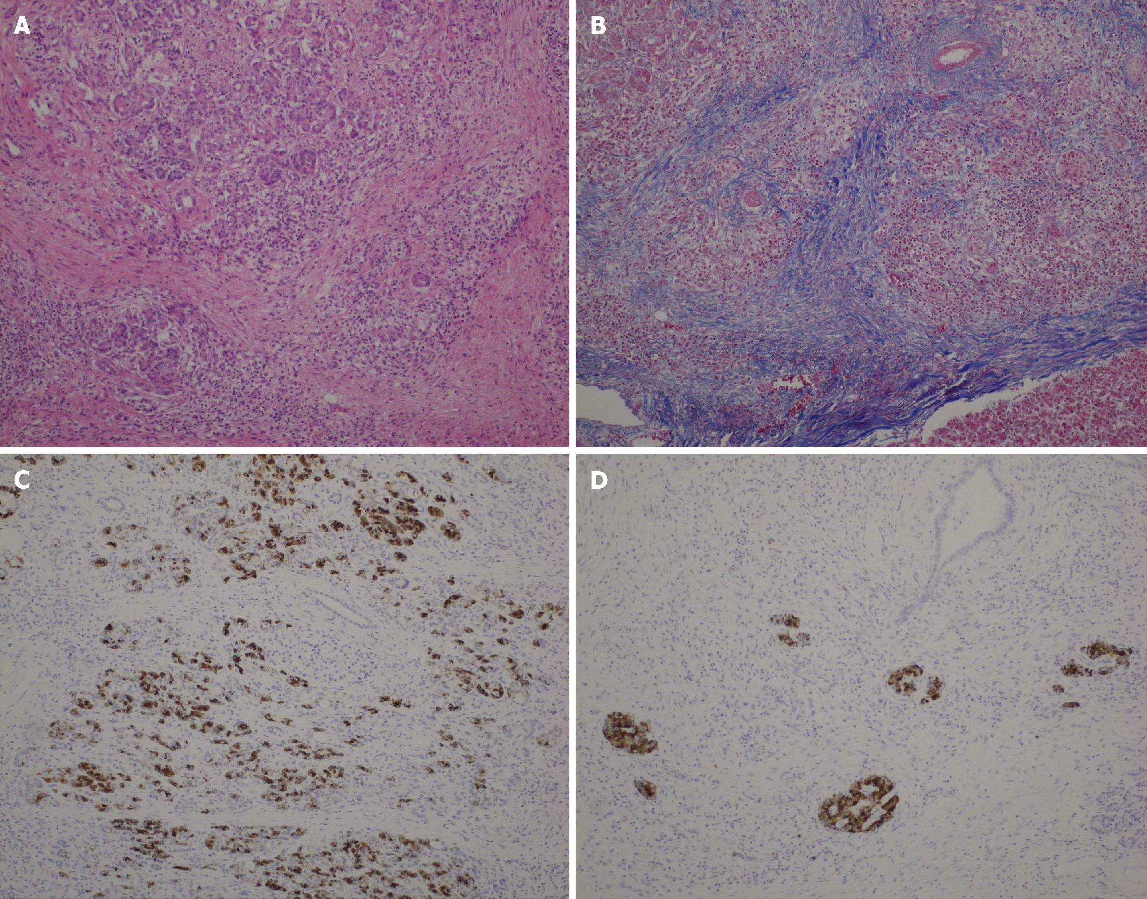
Figure 3 Stained sections from specimens within 1 cm of the pancreatic resection margin.
A: Hematoxylin-eosin staining, 10 × objective lens; B: Masson trichrome staining, 10 × objective lens; C: BCL-10 (333.1) staining, 10 × objective lens; D: Insulin staining, 10 × objective lens.
- Citation: Nakamura A, Ogawa T, Tanaka K, Takahashi Y, Murai S, Tashiro Y, Wada A, Ueda Y, Sasaki Y, Minegishi Y, Matsuo K, Yamochi T. Estimation of pancreatic histology and likelihood of postoperative pancreatic fistula using extracellular volume fraction from contrast-enhanced computed tomography. World J Clin Cases 2025; 13(27): 109243
- URL: https://www.wjgnet.com/2307-8960/full/v13/i27/109243.htm
- DOI: https://dx.doi.org/10.12998/wjcc.v13.i27.109243


