
















©The Author(s) 2025.
World J Clin Cases. Sep 16, 2025; 13(26): 108052
Published online Sep 16, 2025. doi: 10.12998/wjcc.v13.i26.108052
Published online Sep 16, 2025. doi: 10.12998/wjcc.v13.i26.108052
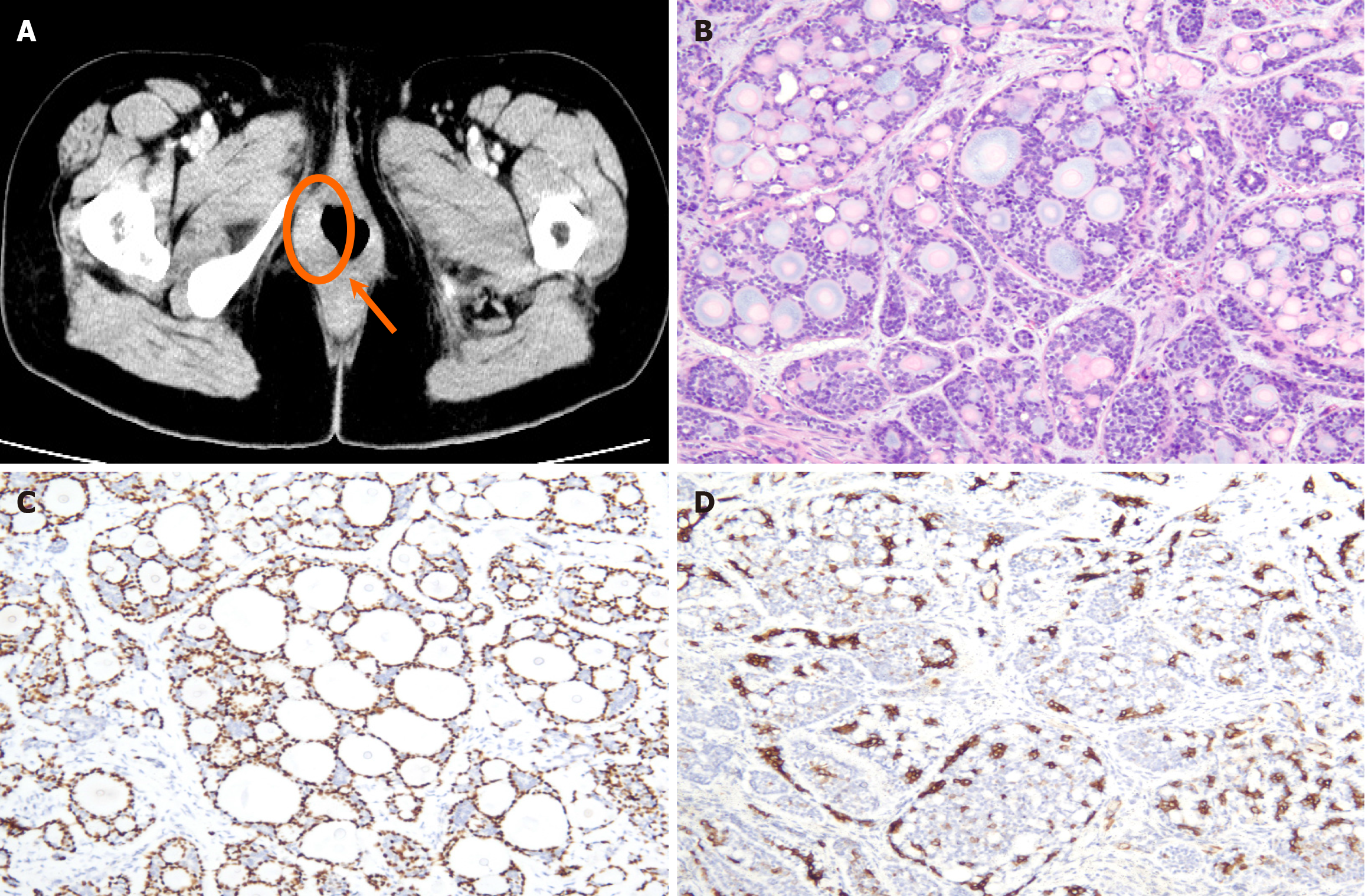
Figure 1 Contrast-enhanced pelvic computed tomography imaging and pathological images of case 1.
A: Pelvic Computed tomography with IV contrast revealed right-side asymmetry in the vulvar region, described as a 1.8 cm × 1.5 cm rounded, uneven reinforcement area with blurred margins; B: Pathological images of hematoxylin and eosin-stained samples showing carcinoma with myoepithelial components. Neoplastic cells with mildly enlarged nuclei were noted, and these cells formed irregular papillary or tubular structure (hematoxylin and eosin × 100); C: P63; D: CK7 revealed diffusely positive staining (EnVision × 100).
- Citation: Liu P, Huang HQ, Bian C, Quan Y. Adenoid cystic carcinoma of the Bartholin’s gland: Two case reports and review of literature. World J Clin Cases 2025; 13(26): 108052
- URL: https://www.wjgnet.com/2307-8960/full/v13/i26/108052.htm
- DOI: https://dx.doi.org/10.12998/wjcc.v13.i26.108052