Copyright
©The Author(s) 2025.
World J Clin Cases. Sep 16, 2025; 13(26): 107748
Published online Sep 16, 2025. doi: 10.12998/wjcc.v13.i26.107748
Published online Sep 16, 2025. doi: 10.12998/wjcc.v13.i26.107748
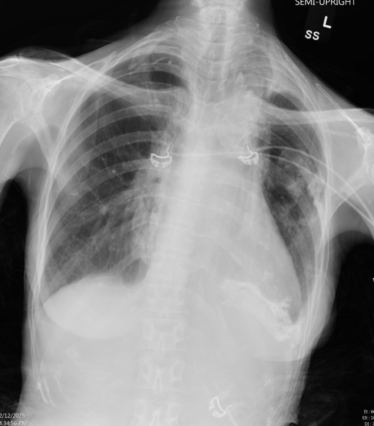
Figure 1 Chest X-ray showing bilateral pleural calcifications and left greater than right apical scarring/posttreatment related changes.
There is also increased focal density within the left lower lung base, suggestive of consolidation.
- Citation: English K, Pick N, Schmitz A. Acute purulent pericarditis secondary to community-acquired streptococcus pneumonia: A case report. World J Clin Cases 2025; 13(26): 107748
- URL: https://www.wjgnet.com/2307-8960/full/v13/i26/107748.htm
- DOI: https://dx.doi.org/10.12998/wjcc.v13.i26.107748


