Copyright
©The Author(s) 2025.
World J Clin Cases. Sep 16, 2025; 13(26): 107496
Published online Sep 16, 2025. doi: 10.12998/wjcc.v13.i26.107496
Published online Sep 16, 2025. doi: 10.12998/wjcc.v13.i26.107496
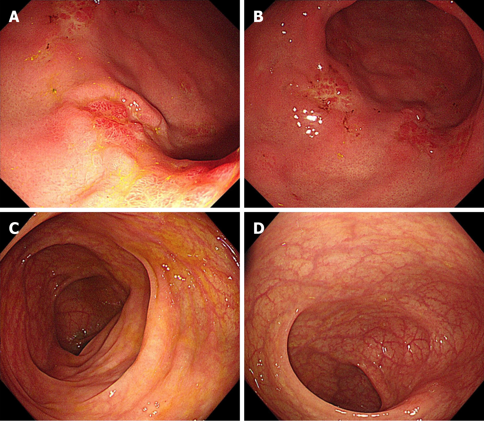
Figure 4 Endoscopic images of upper and lower gastrointestinal tract one and a half years after the detection of gastrointestinal lesions.
A and B: Scattered patchy areas of mucosal hyperplasia are observed in the gastric mucosa, accompanied by focal erosive changes; C and D: The intestinal mucosa appears normal without evidence of pathological lesions.
- Citation: Liu PP, Sun LL, Jing X. Gastrointestinal metastasis from invasive lobular carcinoma following breast cancer treatment: A case report. World J Clin Cases 2025; 13(26): 107496
- URL: https://www.wjgnet.com/2307-8960/full/v13/i26/107496.htm
- DOI: https://dx.doi.org/10.12998/wjcc.v13.i26.107496


