Copyright
©The Author(s) 2025.
World J Clin Cases. Sep 16, 2025; 13(26): 107496
Published online Sep 16, 2025. doi: 10.12998/wjcc.v13.i26.107496
Published online Sep 16, 2025. doi: 10.12998/wjcc.v13.i26.107496
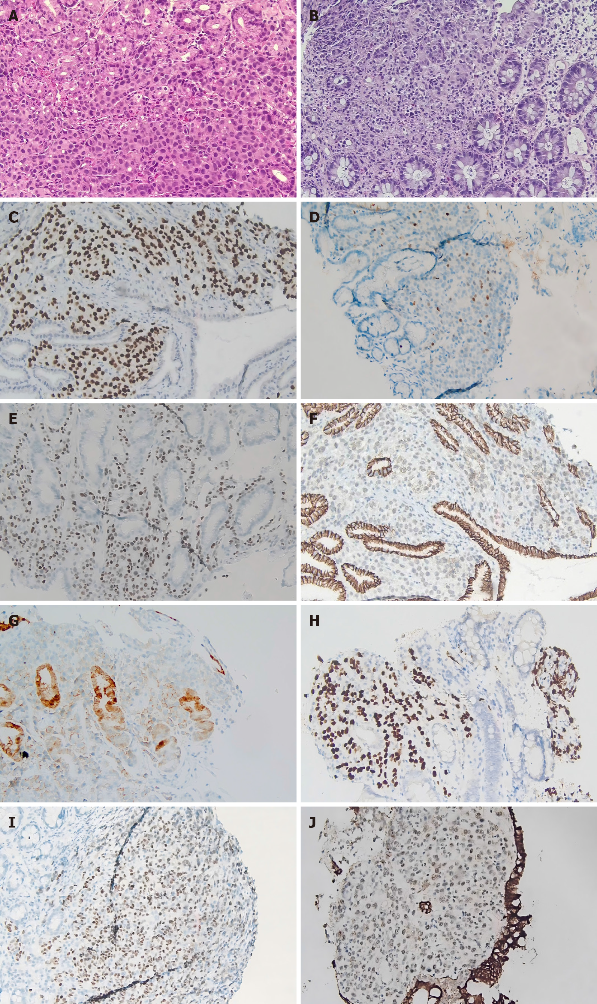
Figure 1 Histopathology and immunohistochemistry findings.
A: Biopsy specimens showing diffuse proliferation of poorly differentiated carcinoma cells in the gastric mucosa [hematoxylin and eosin (HE) staining, 200 ×]; B: Biopsy specimens showing diffuse proliferation of poorly differentiated carcinoma cells in the colonic lamina propria (HE staining, 200 ×); C: Gastric immunohistochemical analysis reveals diffuse expression of GATA-binding protein 3 [immunohistochemistry (IHC) for GATA-3, 200 ×]; D: Gastric immunohistochemical analysis reveals diffuse expression of progesterone receptor(IHC for progesterone receptor, 200 ×); E: Gastric immunohistochemical analysis reveals strong immunoreactivity for estrogen receptor (ER) in about 90% of the neoplastic cells (IHC for ER, 200 ×); F: Gastric immunohistochemical analysis reveals absence of E-cadherin expression (IHC for E-cadherin, 200 ×); G: Human epidermal growth factor receptor 2 (HER2) is faintly stained on the cell membranes of some gastric tumors (IHC for HER2, 200 ×); H: Immunohistochemical staining of colonic lesions shows diffuse expression of GATA-3 (IHC for GATA-3, 100 ×); I: Immunohistochemical staining of colonic lesions shows strong immunoreactivity for ER (IHC for ER, 200 ×); J: Immunohistochemical staining of colonic lesions shows absence of E-cadherin expression (IHC for E-cadherin, 200 ×).
- Citation: Liu PP, Sun LL, Jing X. Gastrointestinal metastasis from invasive lobular carcinoma following breast cancer treatment: A case report. World J Clin Cases 2025; 13(26): 107496
- URL: https://www.wjgnet.com/2307-8960/full/v13/i26/107496.htm
- DOI: https://dx.doi.org/10.12998/wjcc.v13.i26.107496


