Copyright
©The Author(s) 2025.
World J Clin Cases. Sep 16, 2025; 13(26): 107028
Published online Sep 16, 2025. doi: 10.12998/wjcc.v13.i26.107028
Published online Sep 16, 2025. doi: 10.12998/wjcc.v13.i26.107028
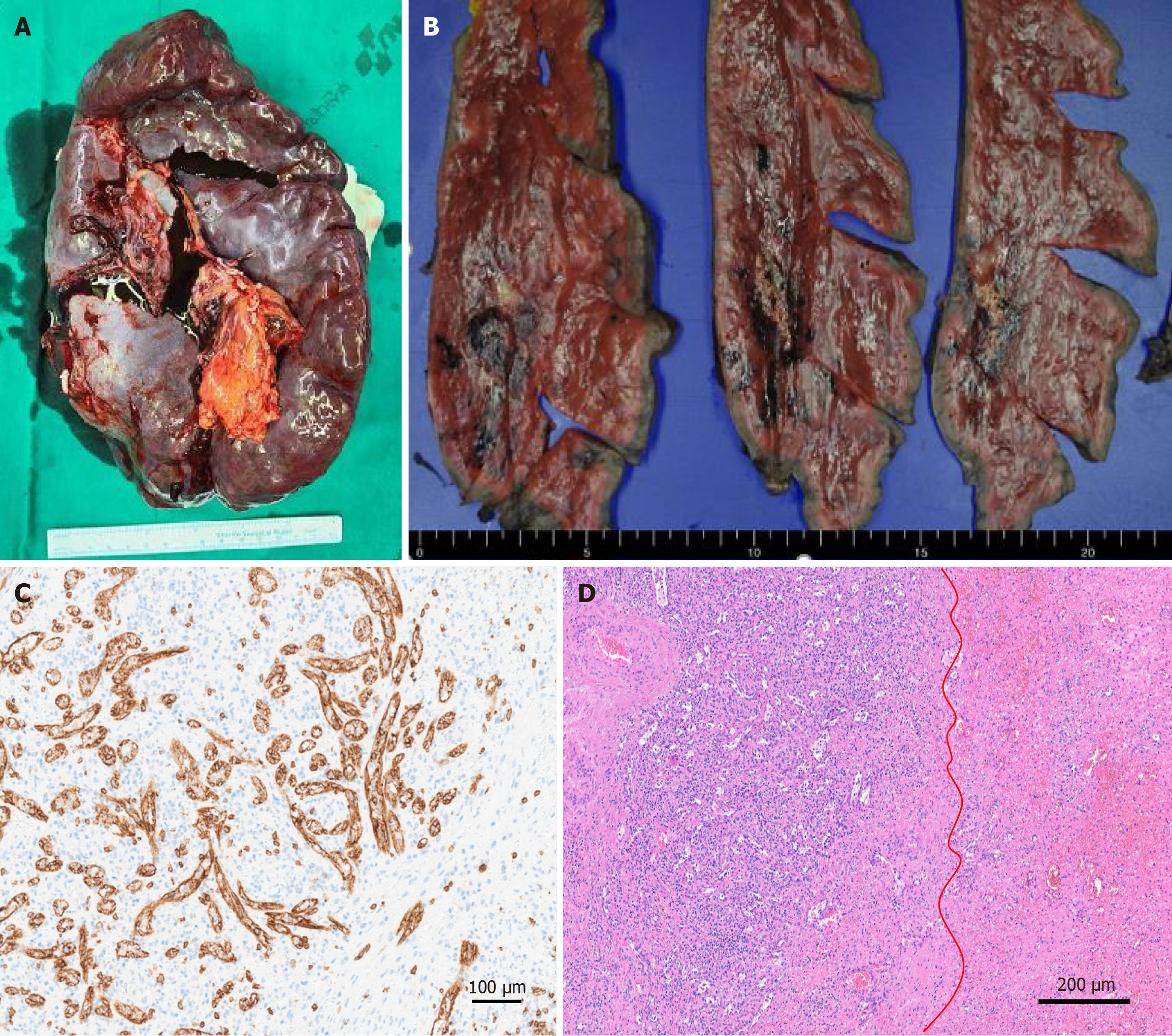
Figure 3 Surgical specimen with histopathological analysis and immunohistochemical examination after splenectomy.
A: The surgical specimen measured 21.7 cm × 15.5 cm × 6.3 cm, with an intact capsule and no signs of congestion; B: Gross examination reveals an ill-defined yellow-white solid mass with hemorrhage, measuring 4.5 cm × 2.5 cm × 2 cm; C: Immunohistochemical staining for CD8+ sinuses (× 200); D: Microscopic findings show vascular proliferation composed of disorganized red pulp elements (left from red line) and hemorrhage (right from red line), visualized with hematoxylin-eosin staining (× 100).
- Citation: Song SB, Noh BG, Oh MH, Yoon M, Park YM, Seo HI, Hong SB, Kim S. Splenic hamartoma mimicking angiosarcoma: A case report. World J Clin Cases 2025; 13(26): 107028
- URL: https://www.wjgnet.com/2307-8960/full/v13/i26/107028.htm
- DOI: https://dx.doi.org/10.12998/wjcc.v13.i26.107028


