Copyright
©The Author(s) 2025.
World J Clin Cases. Sep 16, 2025; 13(26): 107028
Published online Sep 16, 2025. doi: 10.12998/wjcc.v13.i26.107028
Published online Sep 16, 2025. doi: 10.12998/wjcc.v13.i26.107028
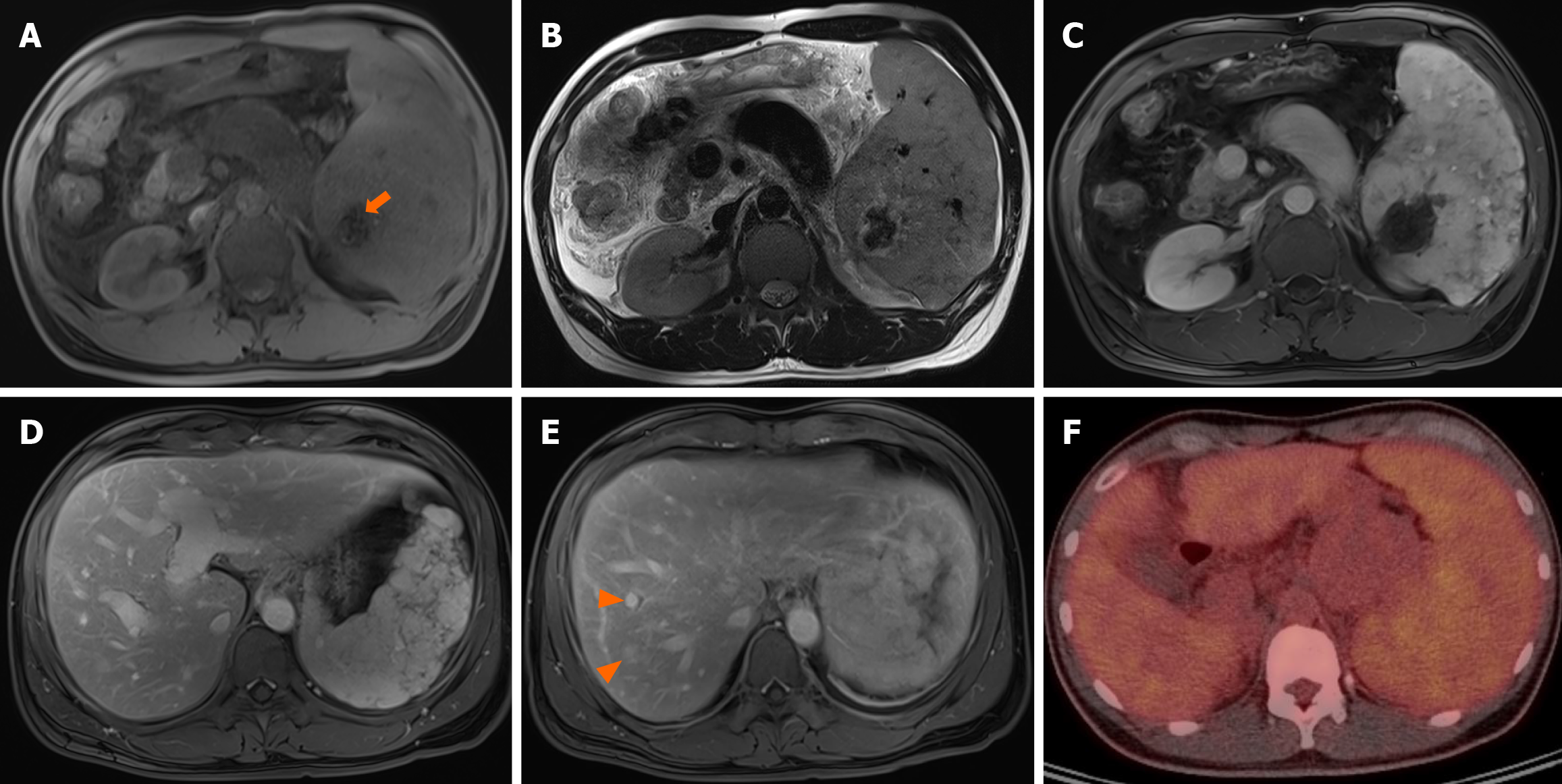
Figure 2 Gadoxetic acid-enhanced liver magnetic resonance imaging and 18F-fluorodeoxyglucose positron emission tomography-computed tomography examination.
The central portion of 8.2 cm × 6.2 cm splenic mass contained hemosiderosis (orange arrow) showing low signal intensity. In dynamic contrast enhanced magnetic resonance images, the splenic mass showed iso to high signal intensity. Contrast enhanced magnetic resonance images demonstrated the multiple hypervascular masses (arrowheads) in liver right hepatic lobe. Positron emission tomography-computed tomography demonstrated splenomegaly with mildly diffusely increased metabolic activity, raising suspicion of hematologic malignancy. A: T1-weighted image; B: T2-weighted image; C-E: In dynamic contrast enhanced magnetic resonance images; F: Positron emission tomography-computed tomography examination image.
- Citation: Song SB, Noh BG, Oh MH, Yoon M, Park YM, Seo HI, Hong SB, Kim S. Splenic hamartoma mimicking angiosarcoma: A case report. World J Clin Cases 2025; 13(26): 107028
- URL: https://www.wjgnet.com/2307-8960/full/v13/i26/107028.htm
- DOI: https://dx.doi.org/10.12998/wjcc.v13.i26.107028


