Copyright
©The Author(s) 2025.
World J Clin Cases. Sep 16, 2025; 13(26): 107028
Published online Sep 16, 2025. doi: 10.12998/wjcc.v13.i26.107028
Published online Sep 16, 2025. doi: 10.12998/wjcc.v13.i26.107028
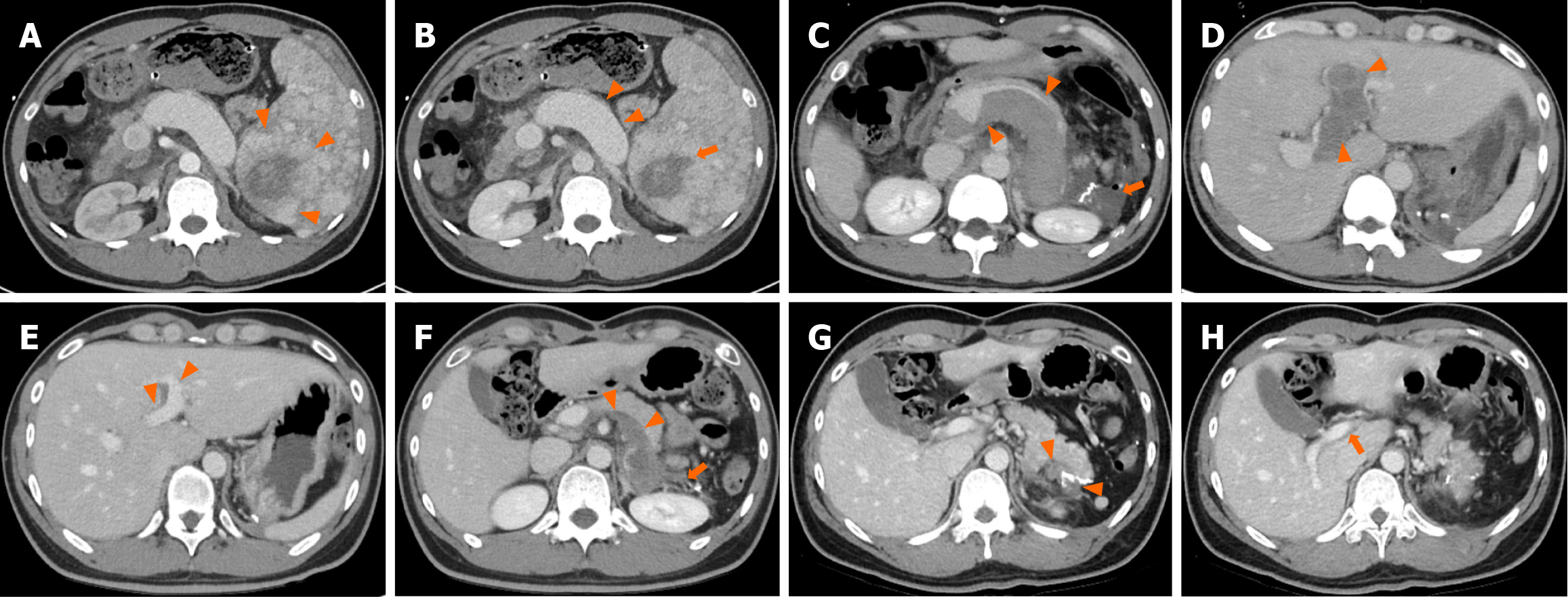
Figure 1 Abdominal contrast-enhanced computed tomography images with postoperative course in a 33-year-old man.
A: Arterial phase contrast-enhanced computed tomography (CT) shows an 8.2 cm × 6.2 cm mass lesion with peripheral rim enhancement (arrowheads); B: Portal venous phase CT reveals a centrally necrotic lesion (orange arrow) with engorged splenic vein (arrowheads); C: On postoperative day 5, portal venous phase CT demonstrates massive splenic vein thrombosis (arrowheads) and a resection site hematoma (orange arrow); D: postoperative day 5, portal venous phase CT also reveals significant intrahepatic and extrahepatic portal vein thrombosis (arrowheads); E: At the 3-month postoperative follow-up, portal venous phase CT shows a remarkable reduction in portal vein thrombosis (arrowheads); F: Significant resolution of remnant splenic vessel thrombosis (arrowheads) and resection site hematoma is observed (orange arrows); G: At the 1-year postoperative follow-up, portal venous phase CT confirms substantial resolution of resection site hematoma (arrowheads); H: Complete resolution of extrahepatic and intrahepatic portal vein thrombosis is seen (orange arrow), with no evidence of recurrence or other abnormalities.
- Citation: Song SB, Noh BG, Oh MH, Yoon M, Park YM, Seo HI, Hong SB, Kim S. Splenic hamartoma mimicking angiosarcoma: A case report. World J Clin Cases 2025; 13(26): 107028
- URL: https://www.wjgnet.com/2307-8960/full/v13/i26/107028.htm
- DOI: https://dx.doi.org/10.12998/wjcc.v13.i26.107028


