
















©The Author(s) 2025.
World J Clin Cases. Sep 16, 2025; 13(26): 105486
Published online Sep 16, 2025. doi: 10.12998/wjcc.v13.i26.105486
Published online Sep 16, 2025. doi: 10.12998/wjcc.v13.i26.105486
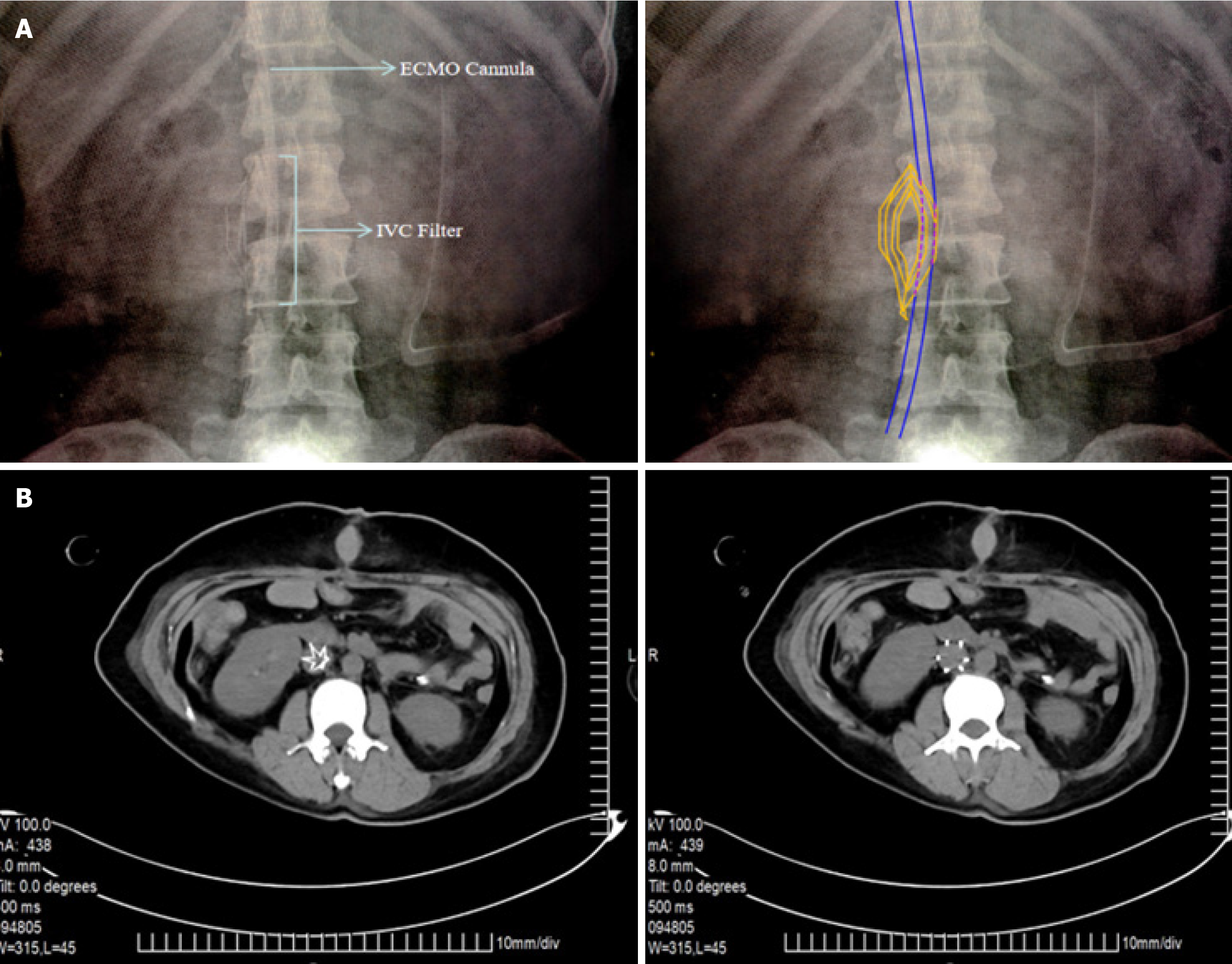
Figure 2 Imaging findings.
A: The extracorporeal membrane oxygenation venous drainage catheter travels in the inferior vena cava through the hollow region of the filter, which is located at the L2 level; B: After extracorporeal membrane oxygenation weaning and removal of the venous drainage catheter, the inferior vena cava filter remained at the L2 level with no thrombus inside or outside the filter.
- Citation: Gao F, Ma S, Xiao X, Yang H, Qian MJ. Emergency veno-arterial extracorporeal membrane oxygenation cannulation through the femoral vein with a pre-positioned inferior vena cava filter: A case report. World J Clin Cases 2025; 13(26): 105486
- URL: https://www.wjgnet.com/2307-8960/full/v13/i26/105486.htm
- DOI: https://dx.doi.org/10.12998/wjcc.v13.i26.105486