Copyright
©The Author(s) 2025.
World J Clin Cases. Sep 16, 2025; 13(26): 104876
Published online Sep 16, 2025. doi: 10.12998/wjcc.v13.i26.104876
Published online Sep 16, 2025. doi: 10.12998/wjcc.v13.i26.104876
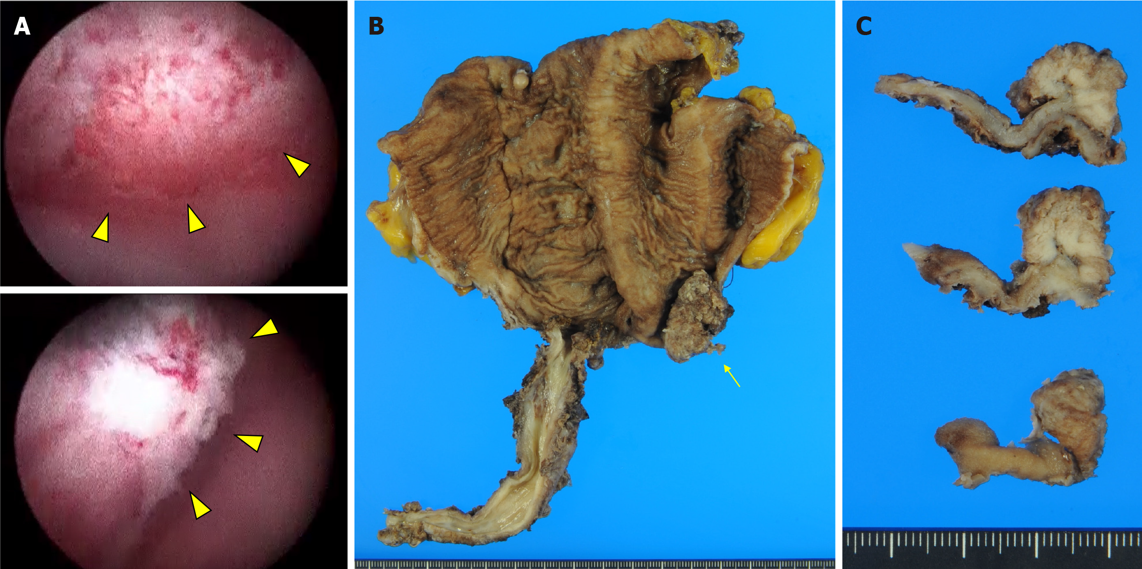
Figure 1 Cystoscopic and macroscopic appearance.
A: Cystoscopy reveals a papillary tumor with white exudate adherent in the left lateral side of the ileal neobladder, which was located distal to the anastomosis (yellow arrowhead); B: Macroscopic appearance of the resected specimen showing an irregular papillary surface with exophytic growth pattern in the ileal neobladder measuring 20 mm × 15 mm. The tumor is located in the left wall of the ileal neobladder (yellow arrow); C: The cut surface of the tumor is grey to white in color.
- Citation: Murakami M, Noguchi H, Matsushita R, Tatarano S, Kirishima M, Tasaki T, Kitazono I, Higashi M, Enokida H, Tanimoto A. Urothelial carcinoma arising in orthotopic ileal neobladder reconstruction: A case report. World J Clin Cases 2025; 13(26): 104876
- URL: https://www.wjgnet.com/2307-8960/full/v13/i26/104876.htm
- DOI: https://dx.doi.org/10.12998/wjcc.v13.i26.104876


