
















©The Author(s) 2025.
World J Clin Cases. Sep 6, 2025; 13(25): 108429
Published online Sep 6, 2025. doi: 10.12998/wjcc.v13.i25.108429
Published online Sep 6, 2025. doi: 10.12998/wjcc.v13.i25.108429
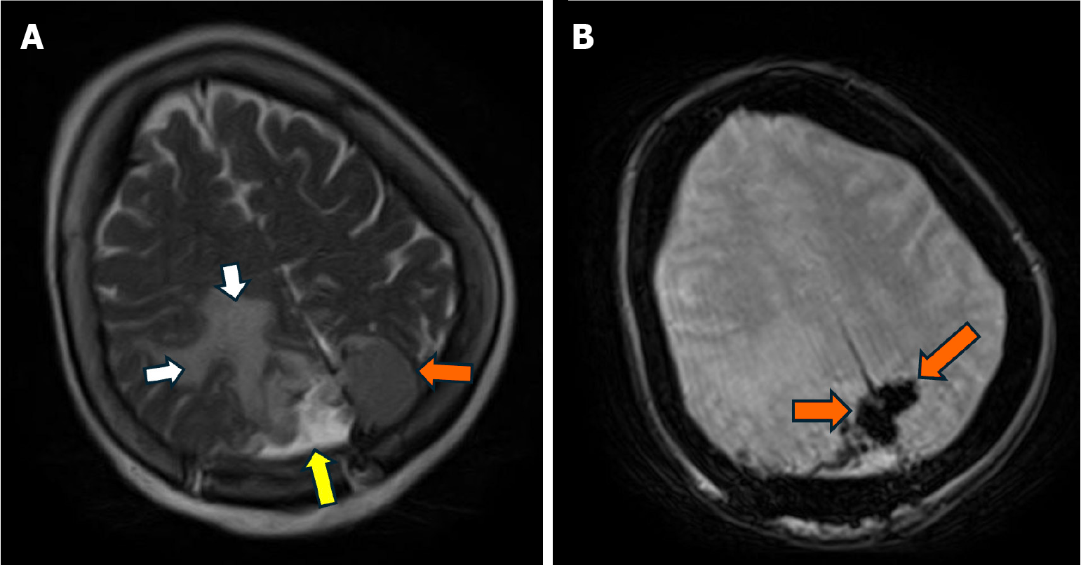
Figure 3 Magnetic resonance imaging.
A and B: Postoperative magnetic resonance imaging brain post-resection with some residual tumor remaining in the left and right posterior parietal parafalcine region (orange arrows). Postoperative blood products and decreased vasogenic edema indicated by yellow and white arrows, respectively.
- Citation: AlSabea N, Syeda S, Gubran M, Gibatova V, Sharma R, Aswani A. Atypical presentation of a large posterior falx meningioma involving the parafalcine region in a 78-year-female: A case report. World J Clin Cases 2025; 13(25): 108429
- URL: https://www.wjgnet.com/2307-8960/full/v13/i25/108429.htm
- DOI: https://dx.doi.org/10.12998/wjcc.v13.i25.108429