Copyright
©The Author(s) 2025.
World J Clin Cases. Sep 6, 2025; 13(25): 108429
Published online Sep 6, 2025. doi: 10.12998/wjcc.v13.i25.108429
Published online Sep 6, 2025. doi: 10.12998/wjcc.v13.i25.108429
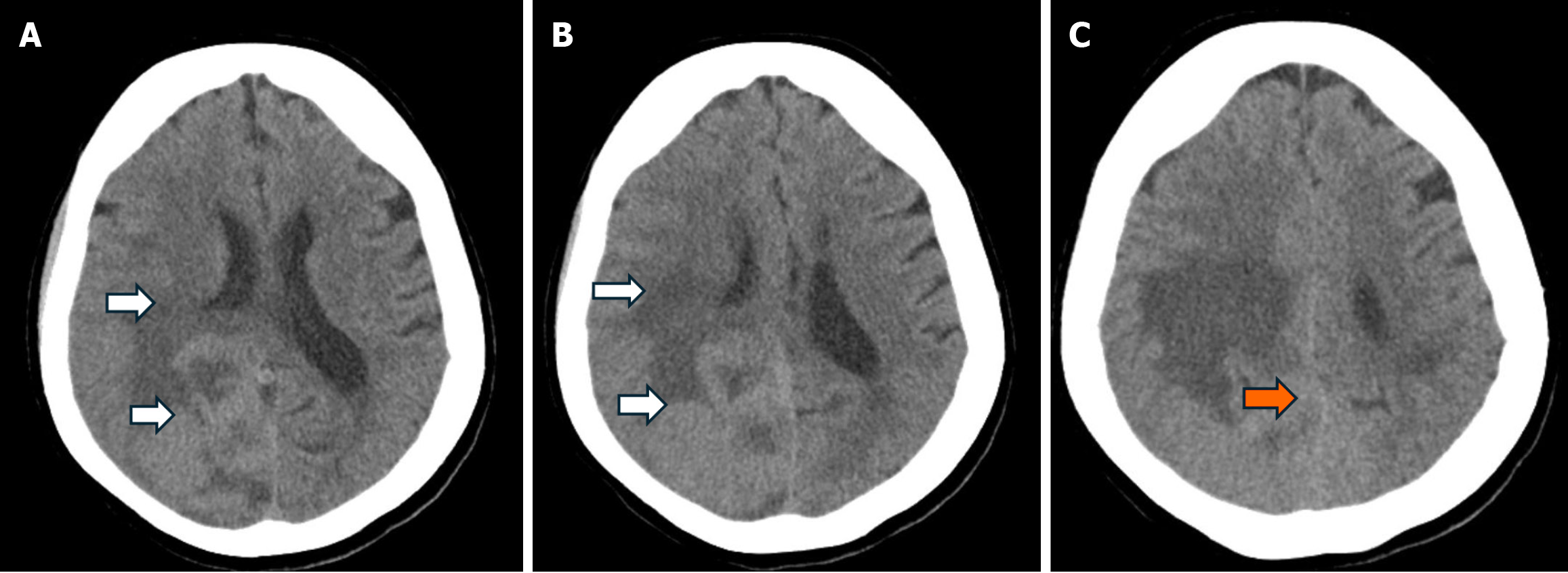
Figure 1 Computed tomography cervical spine, chest, pelvic and knee X-rays were unremarkable.
A-C: Computed tomography head without contrast demonstrating a moderately large area of vasogenic edema (white arrows) seen in the posterior right parietal lobe extending into the right temporal and occipital lobe.
- Citation: AlSabea N, Syeda S, Gubran M, Gibatova V, Sharma R, Aswani A. Atypical presentation of a large posterior falx meningioma involving the parafalcine region in a 78-year-female: A case report. World J Clin Cases 2025; 13(25): 108429
- URL: https://www.wjgnet.com/2307-8960/full/v13/i25/108429.htm
- DOI: https://dx.doi.org/10.12998/wjcc.v13.i25.108429


