Copyright
©The Author(s) 2025.
World J Clin Cases. Sep 6, 2025; 13(25): 107471
Published online Sep 6, 2025. doi: 10.12998/wjcc.v13.i25.107471
Published online Sep 6, 2025. doi: 10.12998/wjcc.v13.i25.107471
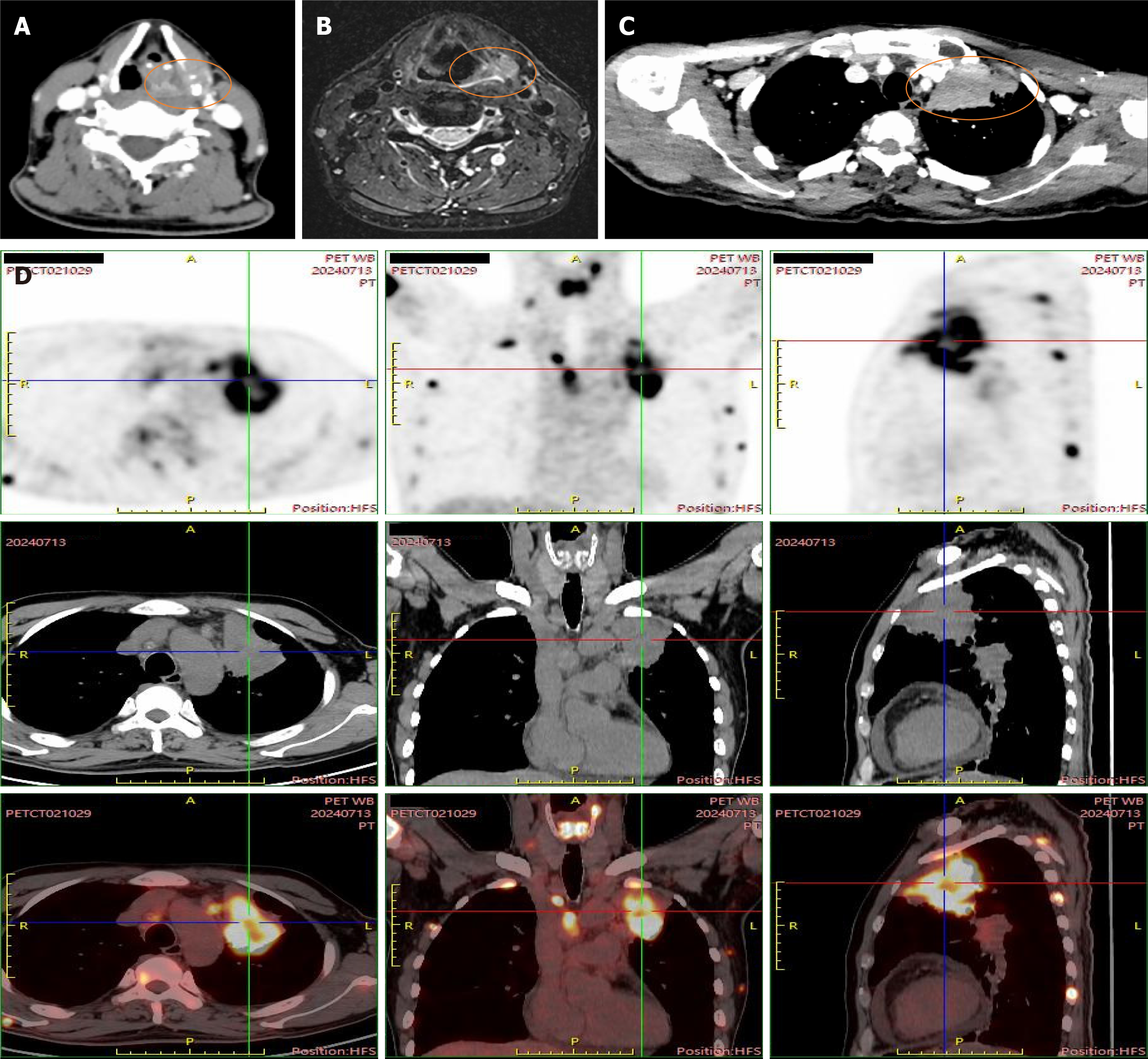
Figure 3 The imaging of the case.
A: Axial computed tomography (CT) with contrast image shows nodules in the left piriform sinus with bone destruction of the thyroid cartilage; B: Magnetic resonance imaging shows nodules in the left piriform sinus; C: CT with contrast image shows peripheral type lung cancer in the left upper lobe; D: Positron emission tomography-CT images. Imaging confirmed widespread metastases involving hilar/mediastinal/supraclavicular/cervical lymph nodes, bilateral adrenals, and multiple bones (spine and thyroid cartilage).
- Citation: Ai MM, Lin T, Guo RY, Zhang YY, Yu F. Unexpected metastasis of thyroid cartilage involvement from lung adenocarcinoma: A case report. World J Clin Cases 2025; 13(25): 107471
- URL: https://www.wjgnet.com/2307-8960/full/v13/i25/107471.htm
- DOI: https://dx.doi.org/10.12998/wjcc.v13.i25.107471


