Copyright
©The Author(s) 2025.
World J Clin Cases. Sep 6, 2025; 13(25): 105028
Published online Sep 6, 2025. doi: 10.12998/wjcc.v13.i25.105028
Published online Sep 6, 2025. doi: 10.12998/wjcc.v13.i25.105028
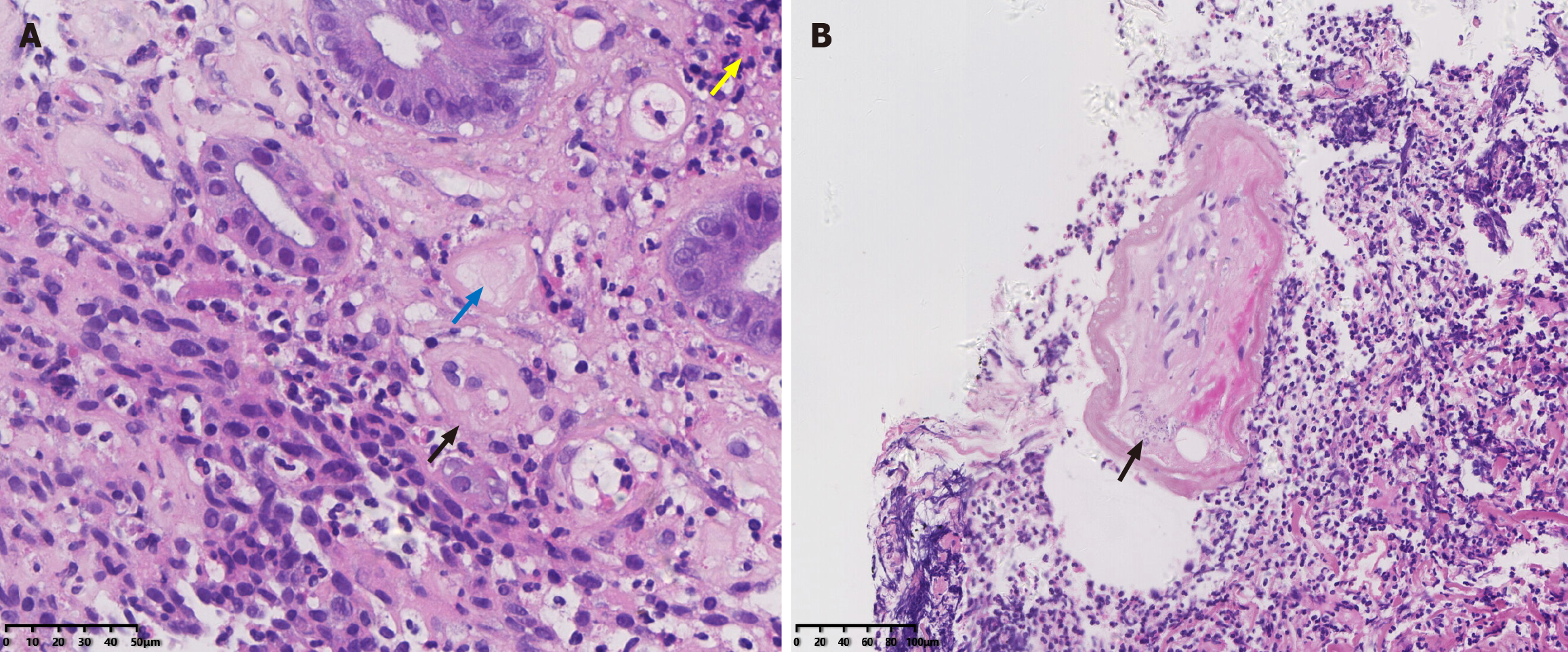
Figure 3 Endoscopic biopsy samples of the left colon.
A: Endoscopic biopsy pathology images showing inflammatory cell infiltration and the deposition of powdery fibrin in the lamina propria (yellow arrow, × 400), fibrous thickening of the wall (black arrow, × 400) of small vessels, a cellulose-like substance (blue arrow, × 400) in the submucosa; B: Calcification surrounding the mesenteric vein (black arrow, × 200).
- Citation: Hou XL, Chen J, Cui MH, Yang GB. Extensive idiopathic mesenteric phlebosclerosis presenting as intestinal pseudo-obstruction: A case report. World J Clin Cases 2025; 13(25): 105028
- URL: https://www.wjgnet.com/2307-8960/full/v13/i25/105028.htm
- DOI: https://dx.doi.org/10.12998/wjcc.v13.i25.105028


