Copyright
©The Author(s) 2025.
World J Clin Cases. Sep 6, 2025; 13(25): 105028
Published online Sep 6, 2025. doi: 10.12998/wjcc.v13.i25.105028
Published online Sep 6, 2025. doi: 10.12998/wjcc.v13.i25.105028
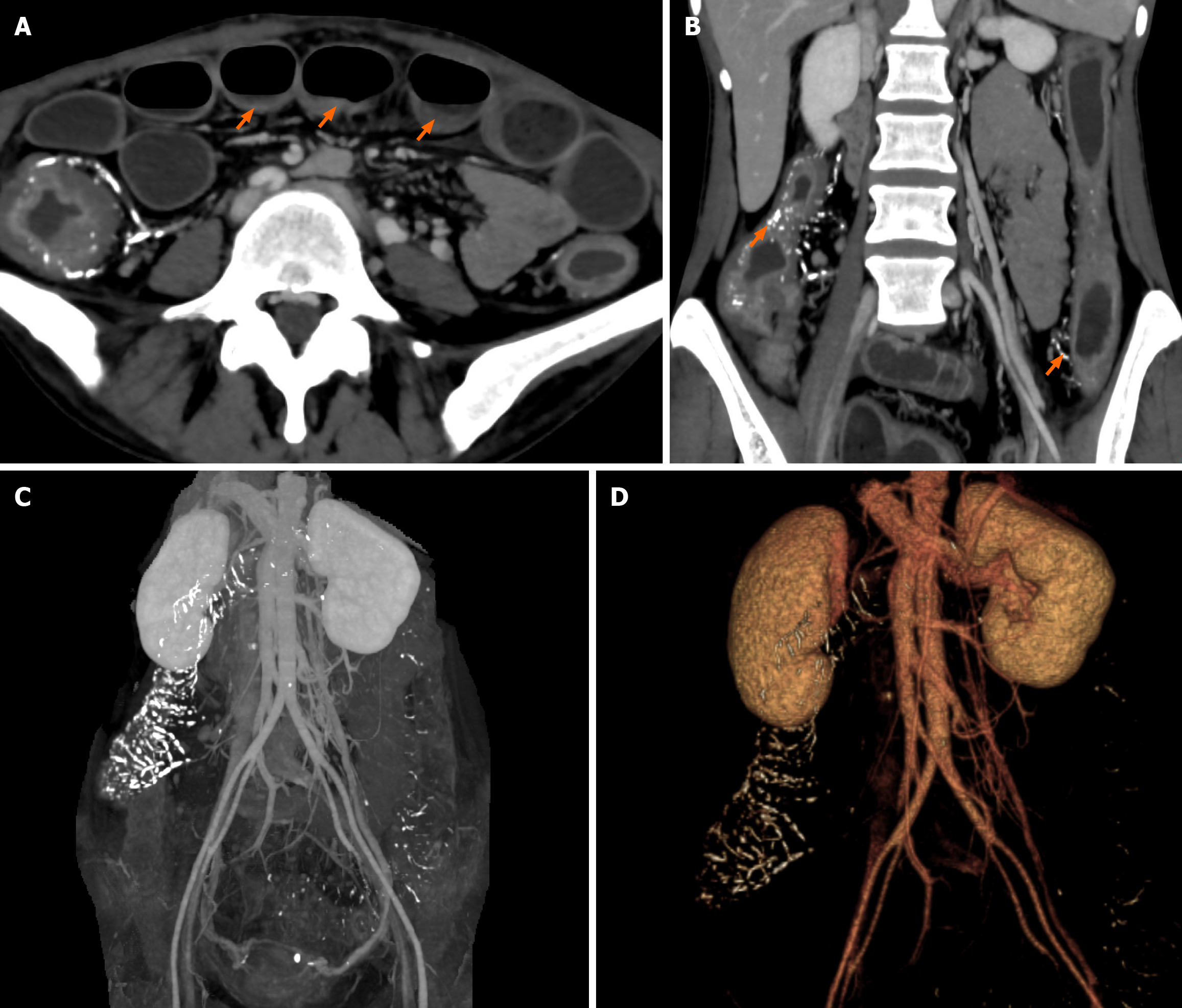
Figure 1 Abdominal computed tomography images revealing remarkable dilatation and fluid collection in the small intestine.
A: Orange arrow, calcification of the pericolonic vein; B: Orange arrow, thickening of the entire colon wall and distal ileum; C and D: Widespread calcification of the distal branches of the superior mesenteric vein and inferior mesenteric vein, among which the calcification of the superior mesenteric vein branches was more severe.
- Citation: Hou XL, Chen J, Cui MH, Yang GB. Extensive idiopathic mesenteric phlebosclerosis presenting as intestinal pseudo-obstruction: A case report. World J Clin Cases 2025; 13(25): 105028
- URL: https://www.wjgnet.com/2307-8960/full/v13/i25/105028.htm
- DOI: https://dx.doi.org/10.12998/wjcc.v13.i25.105028


