Copyright
©The Author(s) 2025.
World J Clin Cases. Aug 26, 2025; 13(24): 107535
Published online Aug 26, 2025. doi: 10.12998/wjcc.v13.i24.107535
Published online Aug 26, 2025. doi: 10.12998/wjcc.v13.i24.107535
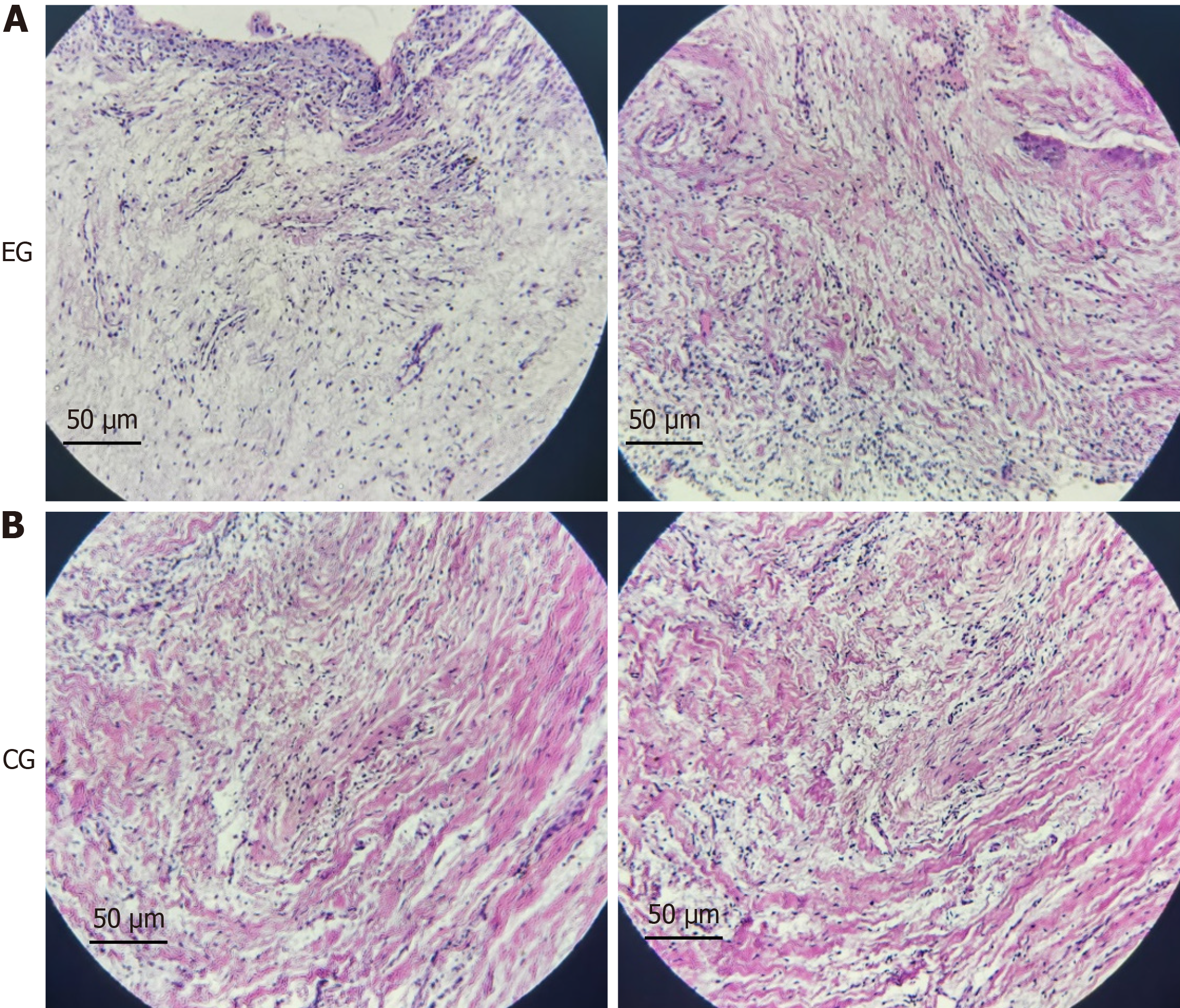
Figure 1 Morphological changes in dental follicles of patients with tooth eruption disorders.
A: The hematoxylin and eosin staining results of dental follicle tissues from the two patients with tooth eruption disorder showed that the volume of dental follicle cells decreased, the nuclei were condensed, and there seemed to be cellular fibrosis; B: The result of hematoxylin and eosin staining of normal dental follicle tissue in the control group (×200).
- Citation: Cai J, Qin H. Mechanism analysis of periostin in osteoclasts differentiation of dental follicle: Two case reports. World J Clin Cases 2025; 13(24): 107535
- URL: https://www.wjgnet.com/2307-8960/full/v13/i24/107535.htm
- DOI: https://dx.doi.org/10.12998/wjcc.v13.i24.107535


