Copyright
©The Author(s) 2025.
World J Clin Cases. Aug 16, 2025; 13(23): 106140
Published online Aug 16, 2025. doi: 10.12998/wjcc.v13.i23.106140
Published online Aug 16, 2025. doi: 10.12998/wjcc.v13.i23.106140
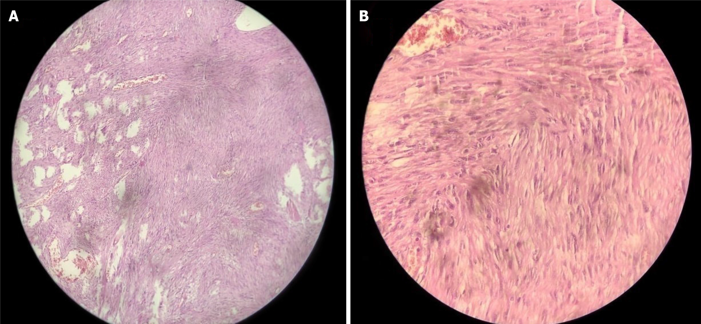
Figure 2 Histopathological image of postoperative jejunal specimen.
A: A 10× view of a submucosal tumor comprising spindle cells arranged in a long, fascicular pattern; B: A 40× view of spindle cells showing minimal pleomorphism mild hyperchromasia, low mitotic index, without any necrosis or epithelioid cells.
- Citation: Maity R, Rathna RB, Dhali A, Fernandes N, Biswas J, Kapoor GS, Dhali GK. Ulcerated benign jejunal gastrointestinal stromal tumor causing gastrointestinal bleeding: A case report. World J Clin Cases 2025; 13(23): 106140
- URL: https://www.wjgnet.com/2307-8960/full/v13/i23/106140.htm
- DOI: https://dx.doi.org/10.12998/wjcc.v13.i23.106140


