Copyright
©The Author(s) 2025.
World J Clin Cases. Aug 16, 2025; 13(23): 106140
Published online Aug 16, 2025. doi: 10.12998/wjcc.v13.i23.106140
Published online Aug 16, 2025. doi: 10.12998/wjcc.v13.i23.106140
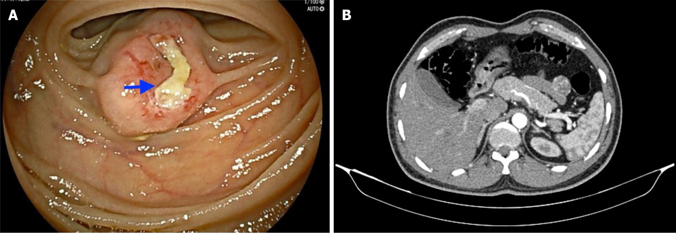
Figure 1 Imaging examinations.
A: Double-balloon enteroscopy image showing a subepithelial lesion (2 cm × 1.5 cm) approximately 150 cm distal to the duodenojejunal flexure and an overlying ulcer without any active bleeding; B: A contrast-enhanced computed tomography abdomen scan revealing a well-defined, round-shaped, homogenously enhancing solid exophytic lesion (30 mm × 22 mm × 26 mm) arising from the proximal jejunal loops without any obvious adjacent organ involvement, internal necrotic foci or calcification.
- Citation: Maity R, Rathna RB, Dhali A, Fernandes N, Biswas J, Kapoor GS, Dhali GK. Ulcerated benign jejunal gastrointestinal stromal tumor causing gastrointestinal bleeding: A case report. World J Clin Cases 2025; 13(23): 106140
- URL: https://www.wjgnet.com/2307-8960/full/v13/i23/106140.htm
- DOI: https://dx.doi.org/10.12998/wjcc.v13.i23.106140


