Copyright
©The Author(s) 2025.
World J Clin Cases. Aug 6, 2025; 13(22): 104283
Published online Aug 6, 2025. doi: 10.12998/wjcc.v13.i22.104283
Published online Aug 6, 2025. doi: 10.12998/wjcc.v13.i22.104283
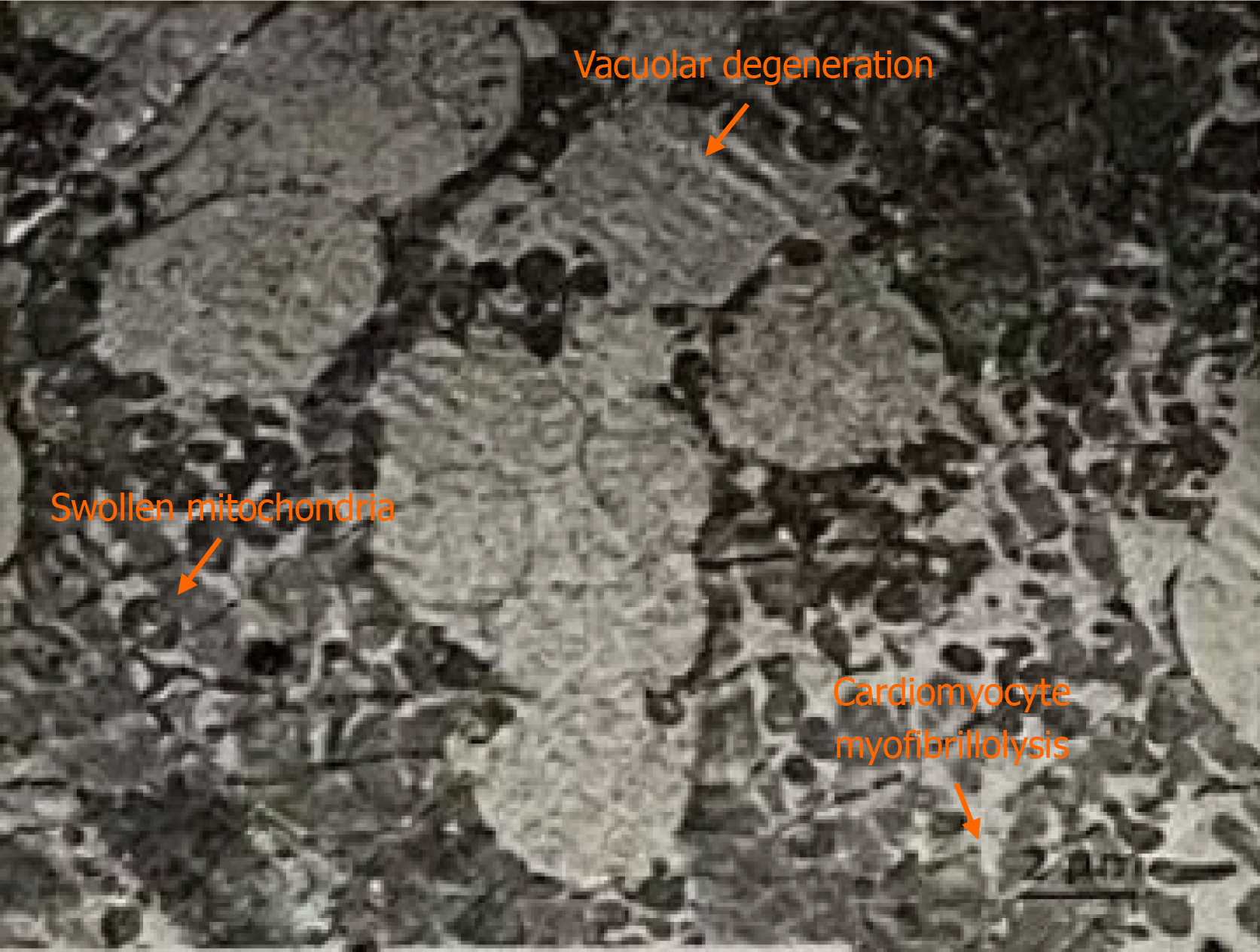
Figure 3 Images from right ventricular septal endocardial myocardial biopsy.
Electron microscopy showed that myofibrils of cardiomyocytes partially dissolved, vacuoles formed in some cells, and electron density increased in some matrices. The mitochondria were swollen slightly but not in large numbers. The basal lamina of the myocardial cells was not clear, and the intercalated disc was not dissected. Many collagen fibers and adipose cells were observed in the interstitium.
- Citation: Xiao PB, Yang XR. Anti-SSA/Ro antibody-positive autoimmune myocarditis combined with complete atrioventricular block requiring implantation with a permanent pacemaker: A case report. World J Clin Cases 2025; 13(22): 104283
- URL: https://www.wjgnet.com/2307-8960/full/v13/i22/104283.htm
- DOI: https://dx.doi.org/10.12998/wjcc.v13.i22.104283


