Copyright
©The Author(s) 2025.
World J Clin Cases. Aug 6, 2025; 13(22): 104283
Published online Aug 6, 2025. doi: 10.12998/wjcc.v13.i22.104283
Published online Aug 6, 2025. doi: 10.12998/wjcc.v13.i22.104283
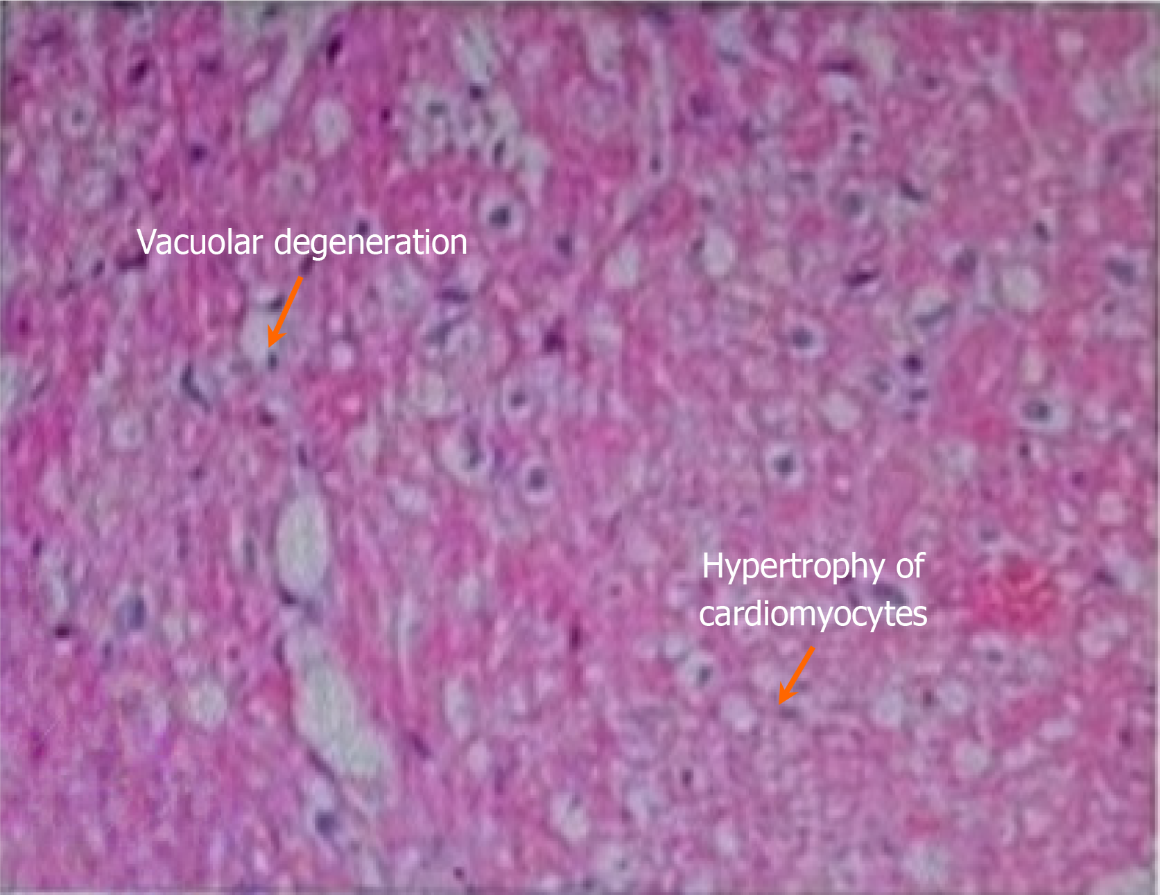
Figure 2 Images from right ventricular septal endocardial myocardial biopsy.
Under the light microscope, slight hypertrophy of cardiomyocytes, vacuolar degeneration, and slight hyperplasia of interstitial fiber tissue were visible.
- Citation: Xiao PB, Yang XR. Anti-SSA/Ro antibody-positive autoimmune myocarditis combined with complete atrioventricular block requiring implantation with a permanent pacemaker: A case report. World J Clin Cases 2025; 13(22): 104283
- URL: https://www.wjgnet.com/2307-8960/full/v13/i22/104283.htm
- DOI: https://dx.doi.org/10.12998/wjcc.v13.i22.104283


