Copyright
©The Author(s) 2025.
World J Clin Cases. Aug 6, 2025; 13(22): 104165
Published online Aug 6, 2025. doi: 10.12998/wjcc.v13.i22.104165
Published online Aug 6, 2025. doi: 10.12998/wjcc.v13.i22.104165
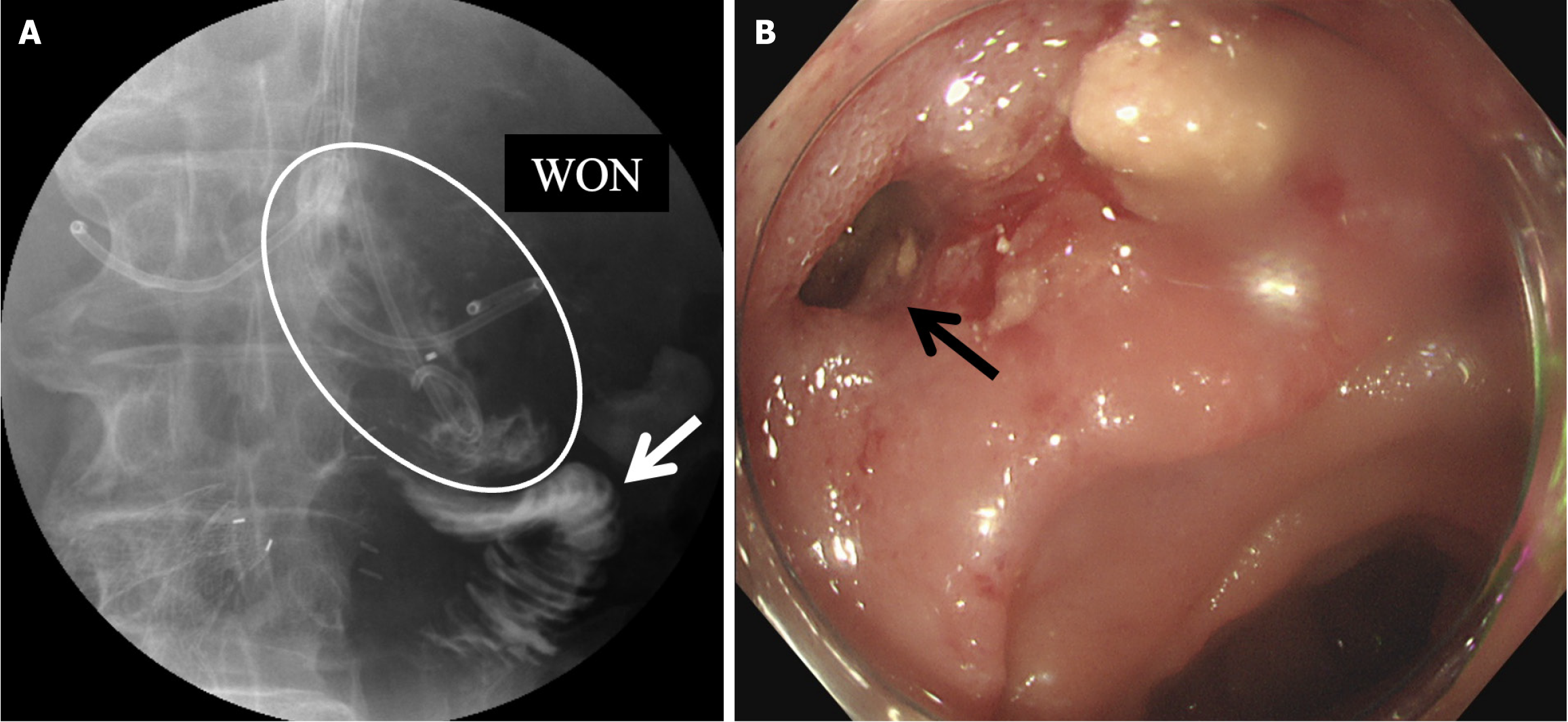
Figure 5 Image examination proved the fistula between walled-off necrosis and the duodenum.
A: The upper gastrointestinal tract contrast study showing a fistula between the walled-off necrosis (white circle) and duodenum (white arrow); B: Endoscopy showing the position of the fistula (black arrow). WON: Walled-off necrosis.
- Citation: Inoue Y, Yata Y, Yokota Y, Li ZL, Kawabata K. Acute pancreatitis after total aortic arch replacement leading to walled-off necrosis: A case report and review of literature. World J Clin Cases 2025; 13(22): 104165
- URL: https://www.wjgnet.com/2307-8960/full/v13/i22/104165.htm
- DOI: https://dx.doi.org/10.12998/wjcc.v13.i22.104165


