Copyright
©The Author(s) 2025.
World J Clin Cases. Jul 26, 2025; 13(21): 105066
Published online Jul 26, 2025. doi: 10.12998/wjcc.v13.i21.105066
Published online Jul 26, 2025. doi: 10.12998/wjcc.v13.i21.105066
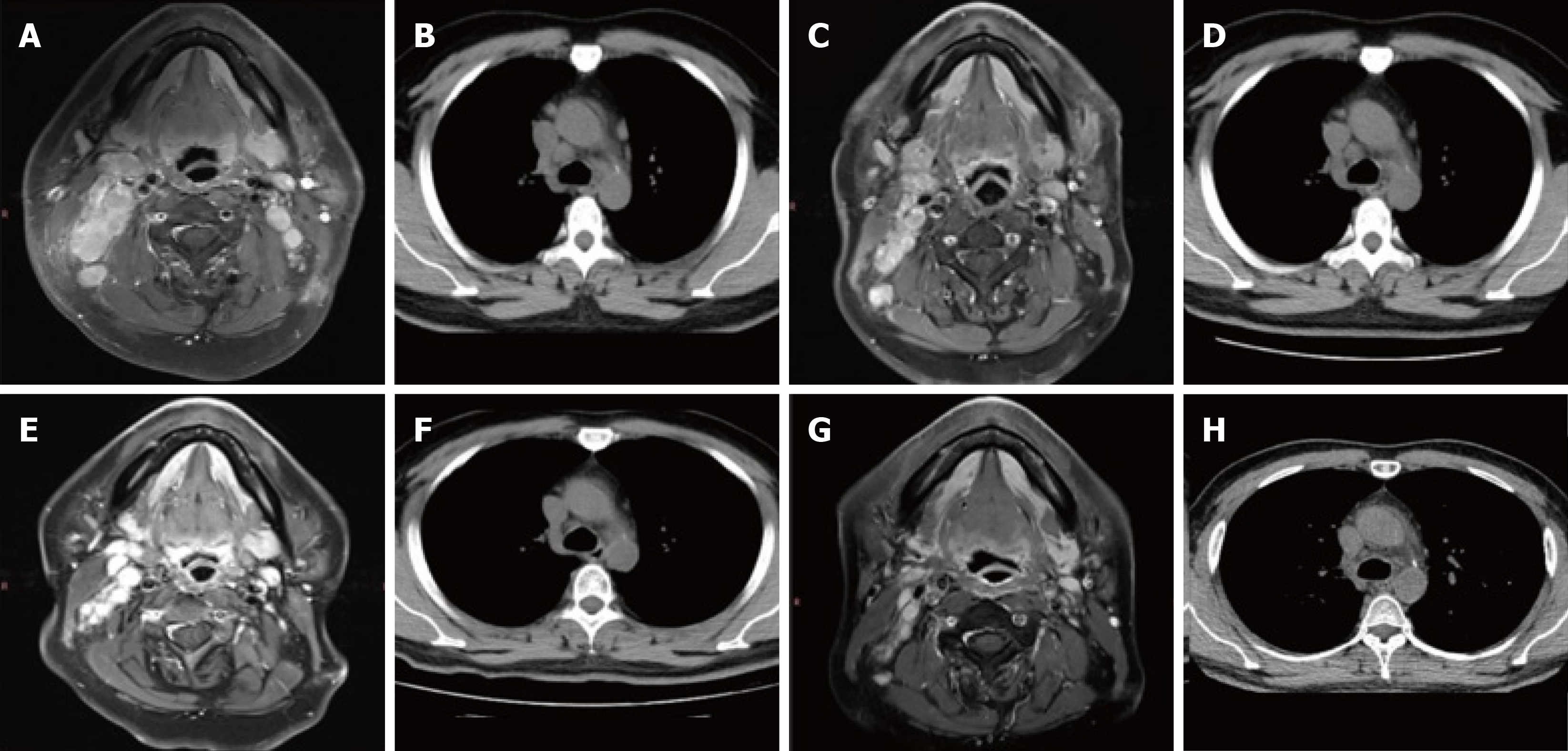
Figure 5 Nasopharynx magnetic resonance imaging (T1+C) and lung computed tomography findings of the nasopharynx.
A: Nasopharynx magnetic resonance imaging (MRI) (July 27, 2022) reveals multiple enlarged lymph nodes in both neck and retropharyngeal groups, with enhancement, partial necrosis, and fusion; B: Lung computed tomography (CT) (August 27, 2022) reveals multiple enlarged lymph nodes in the right neck, bilateral hilum, and mediastinum; C: Nasopharynx MRI (November 03, 2022) reveals that the range of lesions is slightly smaller than that in July; D: Lung CT (December 23, 2022) reveals that bilateral hilar and mediastinal lymph nodes are enlarged; E: Nasopharyngeal MRI (March 8, 2023) reveals that the range of lesions was significantly reduced compared to the October film; F: Lung CT (March 8, 2023) reveals that the range of lesions is significantly reduced compared to the November film; G: Nasopharyngeal MRI (July 5, 2023) reveals swelling and resolution of lymph nodes, diffuse swelling of submandibular soft tissue, slight thickening of the left side of the mucosa in the nasopharynx, and a more uniform signal; H: Lung CT (July 5, 2023) reveals lymph node enlargement subside.
- Citation: Zhou XY, Jiang YJ, Guo XM, Han DH, Liu Y, Qiao Q. Application of circulating tumor DNA liquid biopsy in nasopharyngeal carcinoma: A case report and review of literature. World J Clin Cases 2025; 13(21): 105066
- URL: https://www.wjgnet.com/2307-8960/full/v13/i21/105066.htm
- DOI: https://dx.doi.org/10.12998/wjcc.v13.i21.105066


