
















©The Author(s) 2025.
World J Clin Cases. Jul 26, 2025; 13(21): 103105
Published online Jul 26, 2025. doi: 10.12998/wjcc.v13.i21.103105
Published online Jul 26, 2025. doi: 10.12998/wjcc.v13.i21.103105
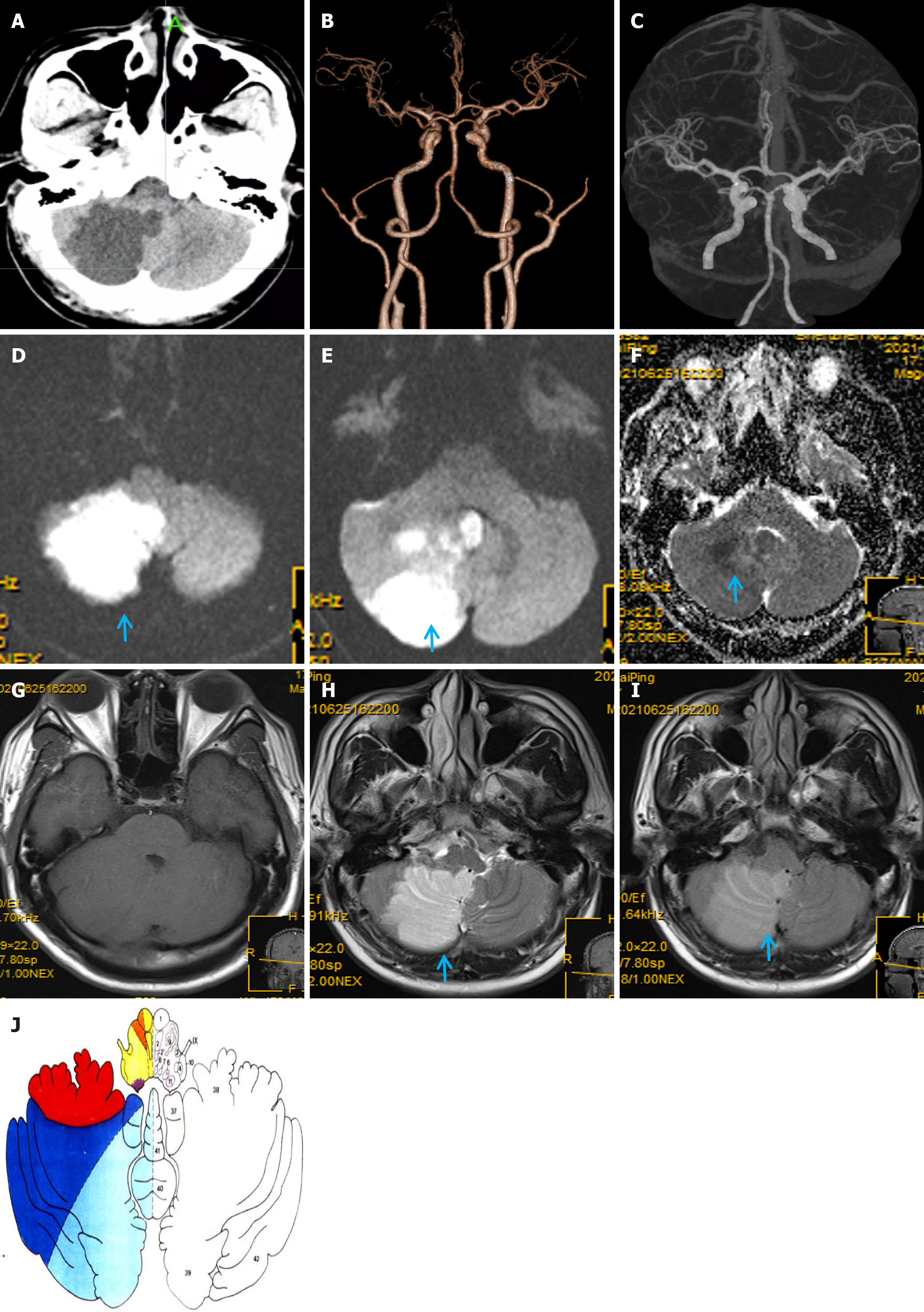
Figure 1 Neuroimaging findings of the patient during hospitalization.
A: Head computed tomography (CT) on admission showing a massive low-density lesion in the right cerebellar hemisphere and vermis; B and C: Craniocervical CT angiography on admission shows no obvious arterial stenosis or occlusion; D-I: Magnetic resonance imaging (MRI) showing a massive acute infarction in the right cerebellar hemisphere and vermis and lesions with limited diffusion on diffusion-weighted imaging (D, E; arrows), hypointensity on the apparent diffusion coefficient map (F, arrow) and T1-weighted imaging (T1WI), and (G) hyperintensity on T2-weighted imaging (H, arrow) and fluid-attenuated inversion recovery MRI (I, arrow); J: Blood supply area of the posterior inferior cerebellar artery (PICA); the dark blue area is the arterial territory of the lateral branch of the PICA, and the light blue area is that of the medial branch.
- Citation: Huang XM, Liao YQ, Cao LM. Massive cerebellar infarction caused by spontaneously isolated posterior inferior cerebellar artery dissection: A case report. World J Clin Cases 2025; 13(21): 103105
- URL: https://www.wjgnet.com/2307-8960/full/v13/i21/103105.htm
- DOI: https://dx.doi.org/10.12998/wjcc.v13.i21.103105