Copyright
©The Author(s) 2025.
World J Clin Cases. Jul 16, 2025; 13(20): 105133
Published online Jul 16, 2025. doi: 10.12998/wjcc.v13.i20.105133
Published online Jul 16, 2025. doi: 10.12998/wjcc.v13.i20.105133
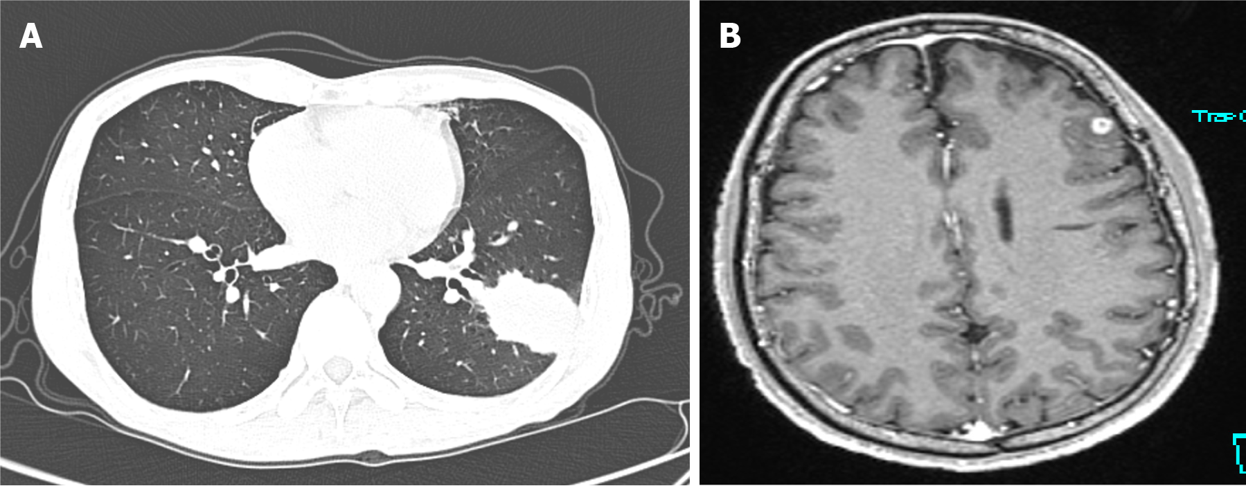
Figure 1 Imaging examinations.
A: Chest computed tomography (December 31, 2020) showing a wedge-shaped high-density lesion in the lower lobe of the left lung; B: Gd-enhanced magnetic resonance imaging (December 2020) showing a small round enhancing lesion in the left frontal lobe.
- Citation: Gao CY, Yang XJ, Guo E, Zheng YL. Invasive pulmonary cryptococcosis mimicking metastatic lung cancer: A case report and review of literature. World J Clin Cases 2025; 13(20): 105133
- URL: https://www.wjgnet.com/2307-8960/full/v13/i20/105133.htm
- DOI: https://dx.doi.org/10.12998/wjcc.v13.i20.105133


