Copyright
©The Author(s) 2025.
World J Clin Cases. Jul 16, 2025; 13(20): 103627
Published online Jul 16, 2025. doi: 10.12998/wjcc.v13.i20.103627
Published online Jul 16, 2025. doi: 10.12998/wjcc.v13.i20.103627
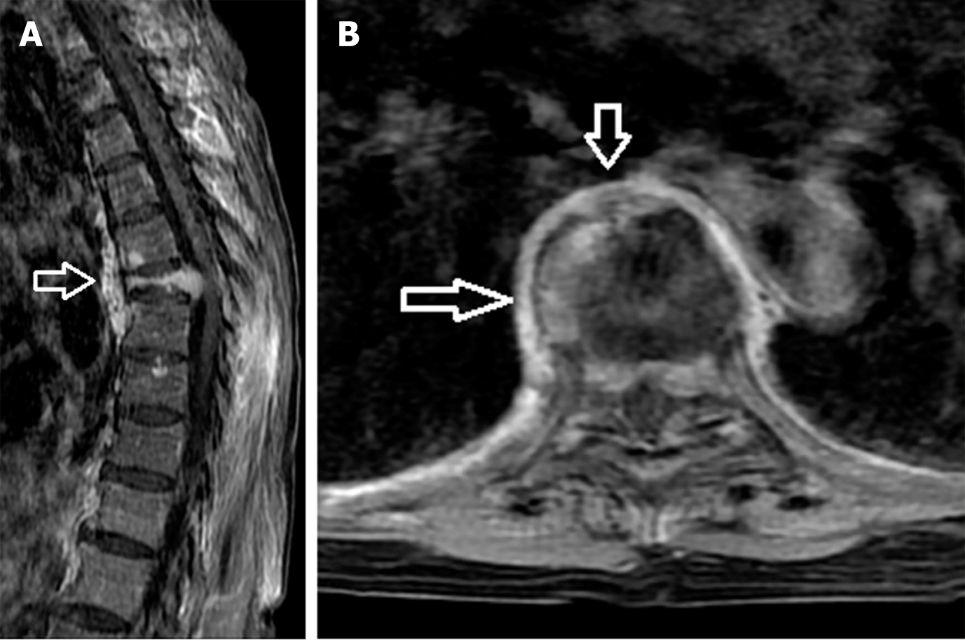
Figure 2 Magnetic resonance images for a 64-year-old woman with multiple myeloma and a malignancy-related compression fracture.
A: Post-enhanced T1-weighted sagittal image showing a hyperintense soft tissue swelling beneath the anterior ligament that has a fusiform appearance and spans multiple vertebral segments (arrow); B: Post-enhanced T1-weighted axial image showing a hyperintense soft tissue swelling with a regular rim-shaped appearance (arrows).
- Citation: Han XL, Shi XL, Li QY, Shao YJ, Gao CP. Paravertebral soft tissue swelling on magnetic resonance images helps in differentiation between osteoporotic and malignant vertebral fractures. World J Clin Cases 2025; 13(20): 103627
- URL: https://www.wjgnet.com/2307-8960/full/v13/i20/103627.htm
- DOI: https://dx.doi.org/10.12998/wjcc.v13.i20.103627


