Copyright
©The Author(s) 2024.
World J Clin Cases. Mar 26, 2024; 12(9): 1685-1690
Published online Mar 26, 2024. doi: 10.12998/wjcc.v12.i9.1685
Published online Mar 26, 2024. doi: 10.12998/wjcc.v12.i9.1685
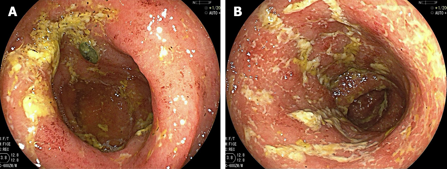
Figure 3 Colonoscopy on December 17, 2022.
A: The mucosa in the sigmoid colon is extensively hyperaemic and oedematous, scattered with multiple irregular shallow ulcers and patchy erosions; B: The hyperaemia and oedema also can be observed in the junction of descending colon and sigmoid colon; and the submucosal vascular texture has disappeared. All the lesions are distributed throughout the mucosa and are covered with a large number of yellow and white secretions.
- Citation: Xu X, Jiang JW, Lu BY, Li XX. Upadacitinib for refractory ulcerative colitis with primary nonresponse to infliximab and vedolizumab: A case report. World J Clin Cases 2024; 12(9): 1685-1690
- URL: https://www.wjgnet.com/2307-8960/full/v12/i9/1685.htm
- DOI: https://dx.doi.org/10.12998/wjcc.v12.i9.1685


