Copyright
©The Author(s) 2024.
World J Clin Cases. Feb 26, 2024; 12(6): 1157-1162
Published online Feb 26, 2024. doi: 10.12998/wjcc.v12.i6.1157
Published online Feb 26, 2024. doi: 10.12998/wjcc.v12.i6.1157
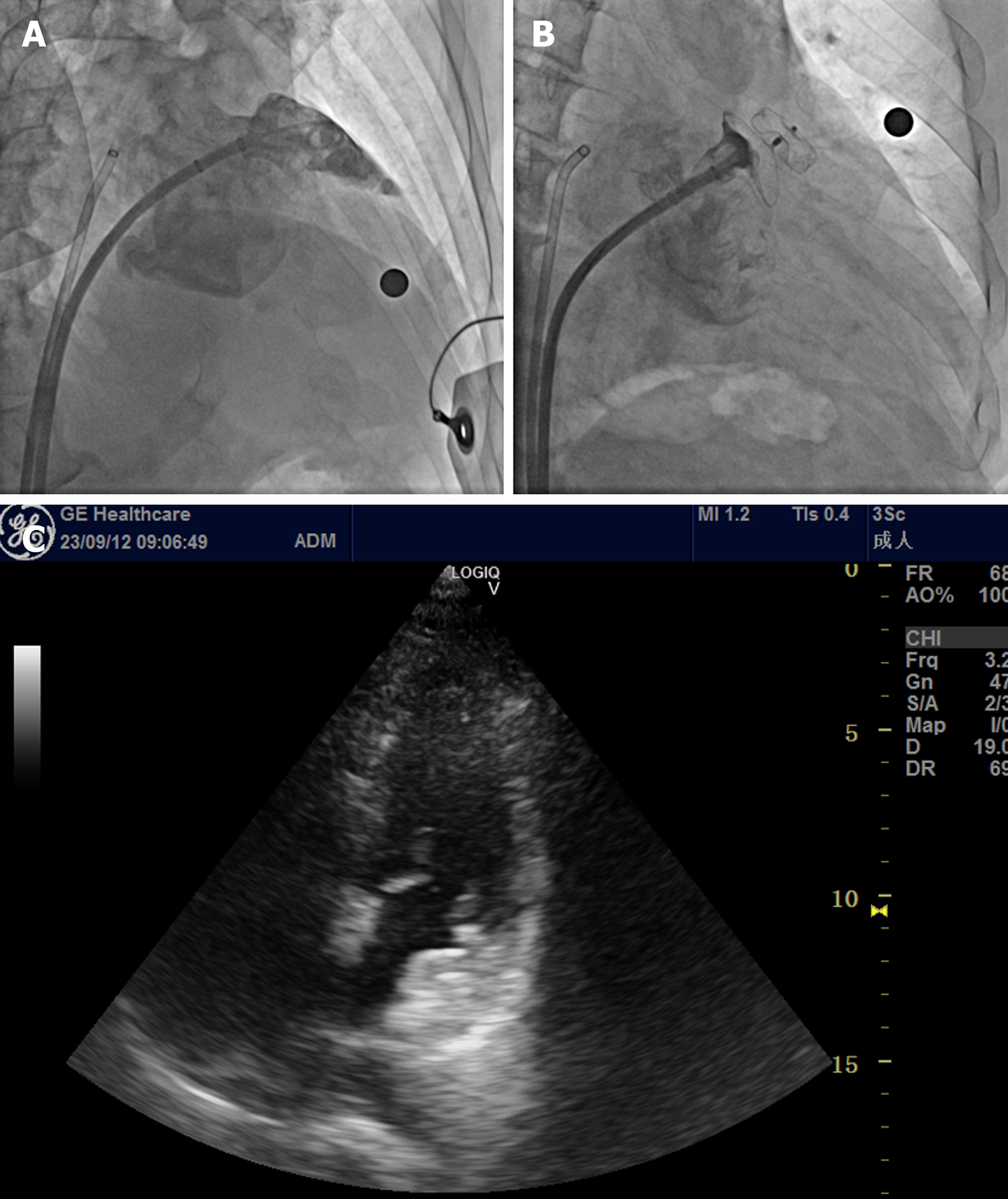
Figure 1 First surgery: Left atrial appendage occlusion.
A: Left atrial appendage angiography before occlusion; B: Before the release of LACbes 22 mm × 32 mm occluder, no leakage of contrast agent was obvious around the umbrella during the imaging examination; C: Transthoracic echocardiography shows that the occluder is fixed to the opening of the left atrial appendage, and no blood flow signal is observed from the left atrial appendage or left atrium around the umbrella.
- Citation: Yu K, Mei YH. Left atrial appendage occluder detachment treated with transthoracic ultrasound combined with digital subtraction angiography guided catcher: A case report. World J Clin Cases 2024; 12(6): 1157-1162
- URL: https://www.wjgnet.com/2307-8960/full/v12/i6/1157.htm
- DOI: https://dx.doi.org/10.12998/wjcc.v12.i6.1157


