Copyright
©The Author(s) 2024.
World J Clin Cases. Feb 26, 2024; 12(6): 1130-1137
Published online Feb 26, 2024. doi: 10.12998/wjcc.v12.i6.1130
Published online Feb 26, 2024. doi: 10.12998/wjcc.v12.i6.1130
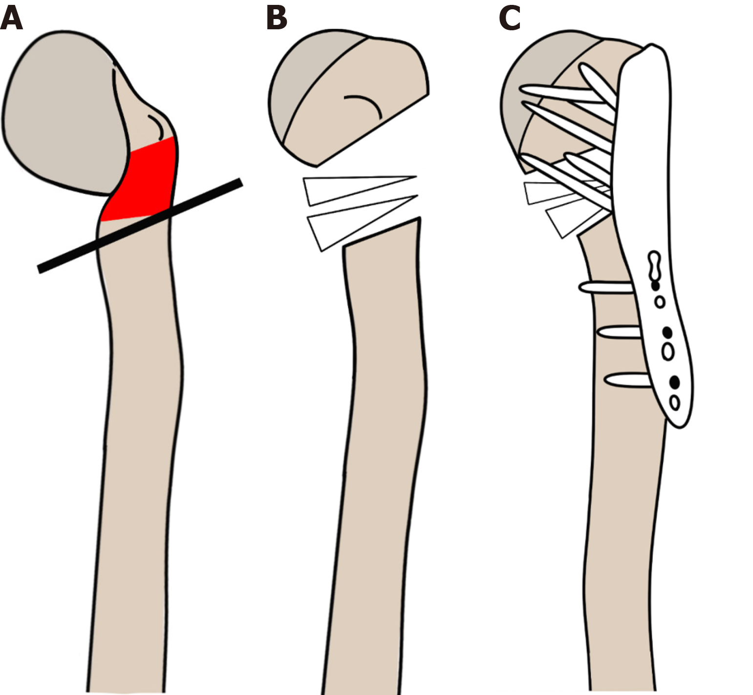
Figure 3 Surgical procedure for acute correction of the proximal humerus for the varus deformity.
The red area indicates the location of the simple bone cyst (SBC). A: Osteotomy was performed in the most angulated area, removing the deformity site of the SBC; B: The gap was filled with allograft (donor femoral head); C: The proximal humerus was fixed with an anatomical locking plate (Depuy-Synthesis®, Raynham, MA, United States) in a valgus position.
- Citation: Lin CS, Lin SM, Rwei SP, Chen CW, Lan TY. Simple bone cysts of the proximal humerus presented with limb length discrepancy: A case report. World J Clin Cases 2024; 12(6): 1130-1137
- URL: https://www.wjgnet.com/2307-8960/full/v12/i6/1130.htm
- DOI: https://dx.doi.org/10.12998/wjcc.v12.i6.1130


