
















©The Author(s) 2024.
World J Clin Cases. Dec 6, 2024; 12(34): 6728-6735
Published online Dec 6, 2024. doi: 10.12998/wjcc.v12.i34.6728
Published online Dec 6, 2024. doi: 10.12998/wjcc.v12.i34.6728
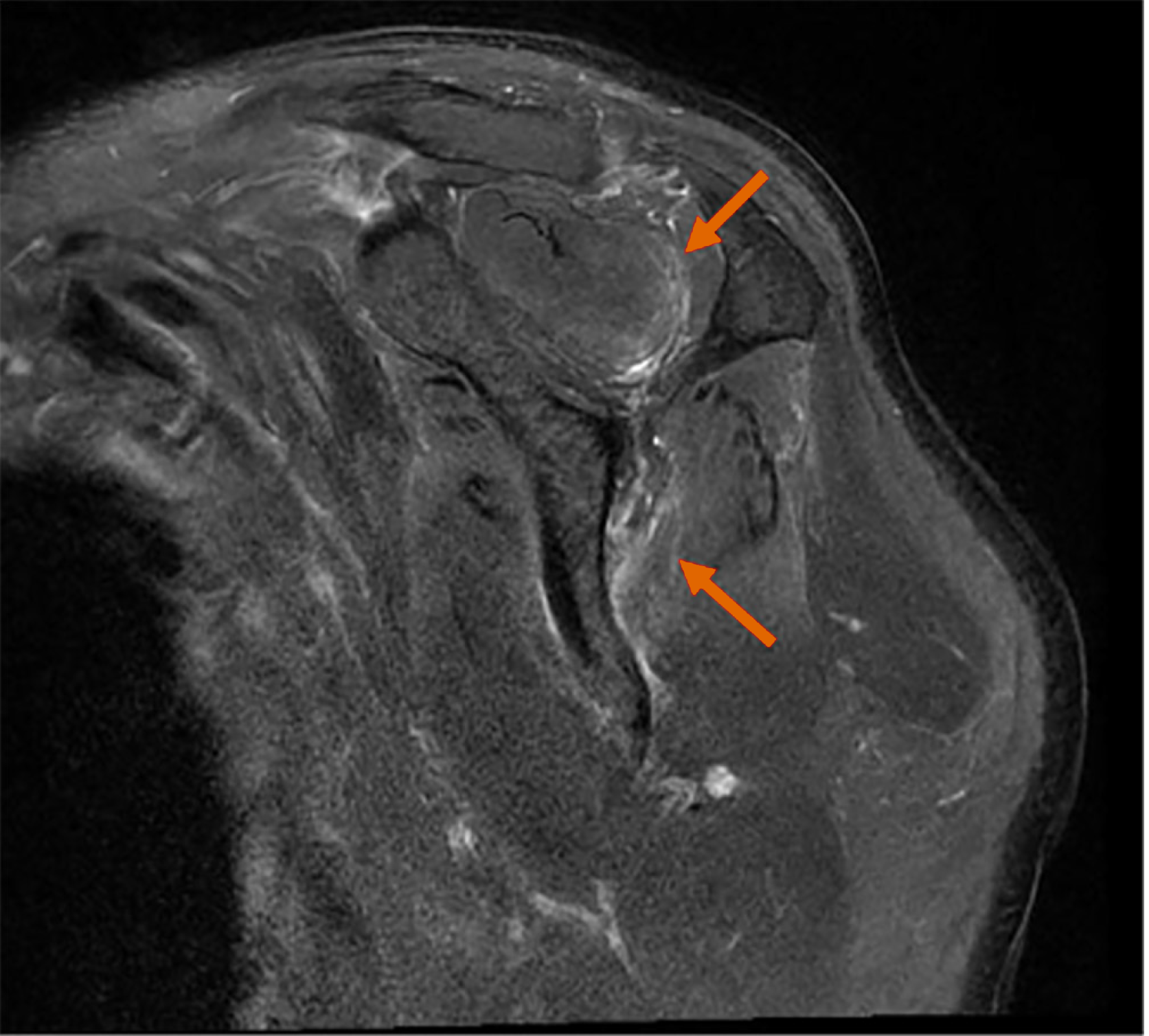
Figure 3 T2-weighted fat saturated oblique sagittal image of shoulder magnetic resonance imaging.
Increased T2 signal intensities of both supraspinatus (upper arrow) and infraspinatus (lower arrow) muscles are noted.
- Citation: Bang MH, Song HL, Hahn S, Kim W, Do HK. Neuralgic amyotrophy with hourglass-like constrictions: A case report. World J Clin Cases 2024; 12(34): 6728-6735
- URL: https://www.wjgnet.com/2307-8960/full/v12/i34/6728.htm
- DOI: https://dx.doi.org/10.12998/wjcc.v12.i34.6728