
















©The Author(s) 2024.
World J Clin Cases. Dec 6, 2024; 12(34): 6705-6714
Published online Dec 6, 2024. doi: 10.12998/wjcc.v12.i34.6705
Published online Dec 6, 2024. doi: 10.12998/wjcc.v12.i34.6705
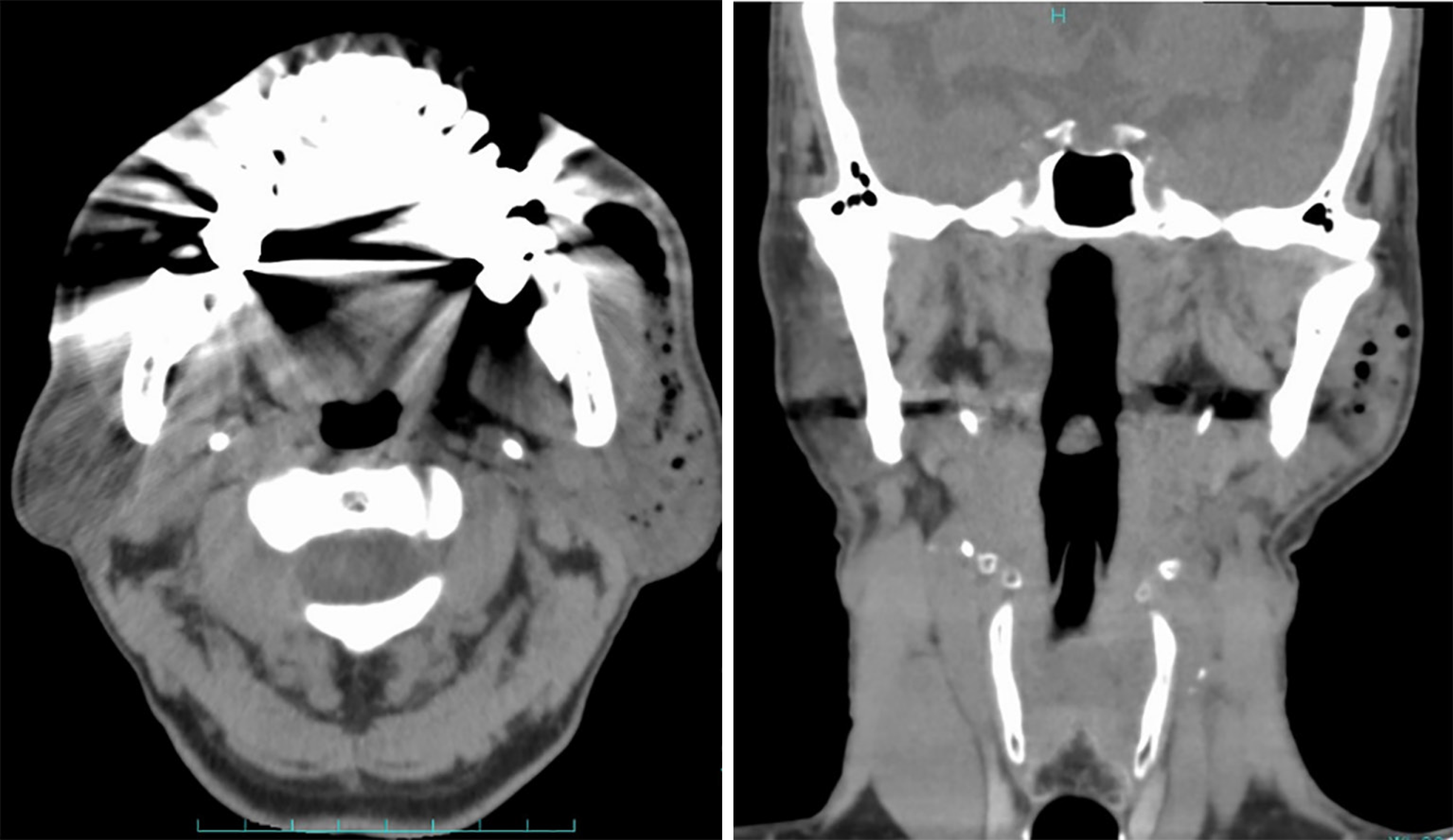
Figure 3 Computed tomography scans reveal swelling of the left cheek.
Marked dilation with air density is observed extending from the left Stensen’s duct to the intraparotid duct. No subcutaneous emphysema is noted.
- Citation: Kubota W, Kyan-Onodera M, Fujimoto Y, Sakuma A, Katada R, Sugiura C. Pneumoparotid with imaging findings: A case report and review of literature. World J Clin Cases 2024; 12(34): 6705-6714
- URL: https://www.wjgnet.com/2307-8960/full/v12/i34/6705.htm
- DOI: https://dx.doi.org/10.12998/wjcc.v12.i34.6705