















©The Author(s) 2024.
World J Clin Cases. Nov 16, 2024; 12(32): 6551-6558
Published online Nov 16, 2024. doi: 10.12998/wjcc.v12.i32.6551
Published online Nov 16, 2024. doi: 10.12998/wjcc.v12.i32.6551
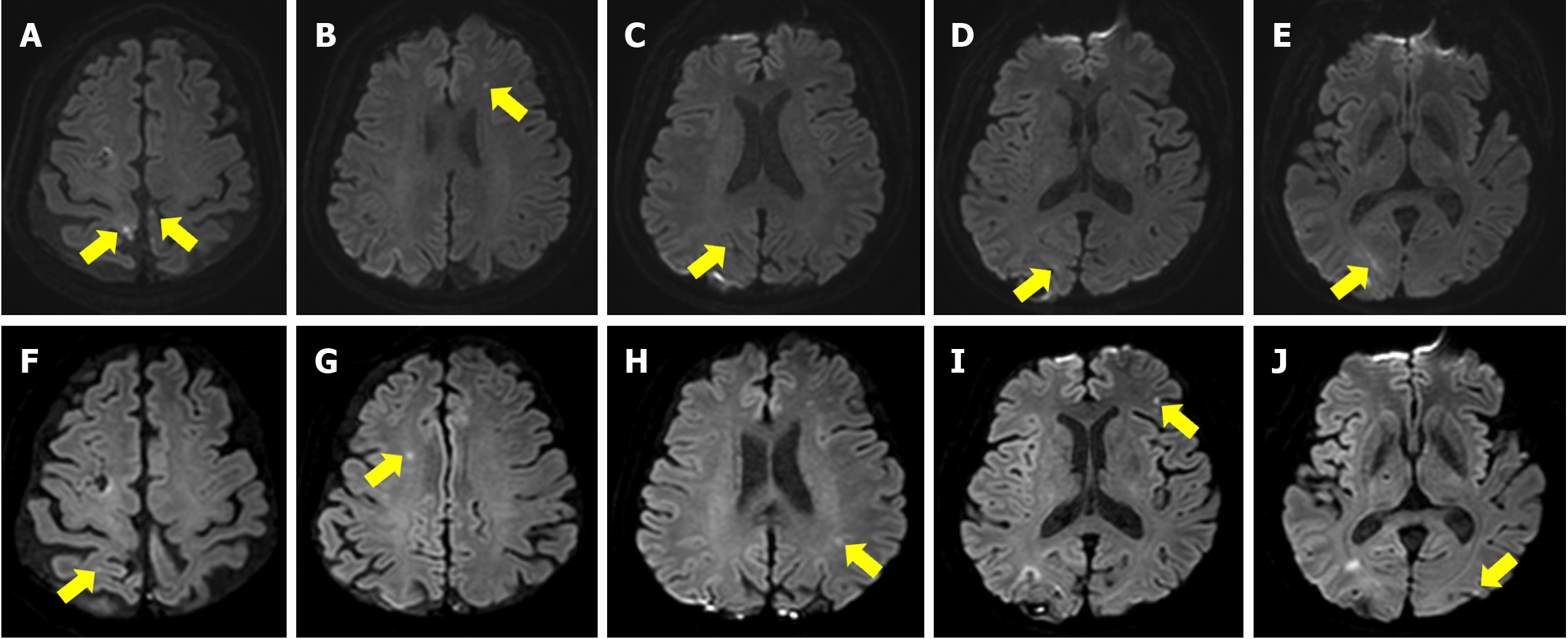
Figure 1 Brain magnetic resonance imaging on hospital day 1 and follow-up brain magnetic resonance imaging on hospital day 19.
A-E: Brain magnetic resonance imaging on hospital day 1. Diffusion weighted image showing focal infarction in the bilateral parietal lobes (A), left frontal lobe (B), right occipital lobe (C-E); F-J: Follow-up brain magnetic resonance imaging on hospital day 19. Right parietal lobe (F and G), left parietal lobe (H), left frontal lobe (I), left occipital lobe (J).
- Citation: Chae TS, Kim DS, Kim GW, Won YH, Ko MH, Park SH, Seo JH. Immunoglobulin G4-related spinal pachymeningitis: A case report. World J Clin Cases 2024; 12(32): 6551-6558
- URL: https://www.wjgnet.com/2307-8960/full/v12/i32/6551.htm
- DOI: https://dx.doi.org/10.12998/wjcc.v12.i32.6551