Copyright
©The Author(s) 2024.
World J Clin Cases. Jan 26, 2024; 12(3): 616-622
Published online Jan 26, 2024. doi: 10.12998/wjcc.v12.i3.616
Published online Jan 26, 2024. doi: 10.12998/wjcc.v12.i3.616
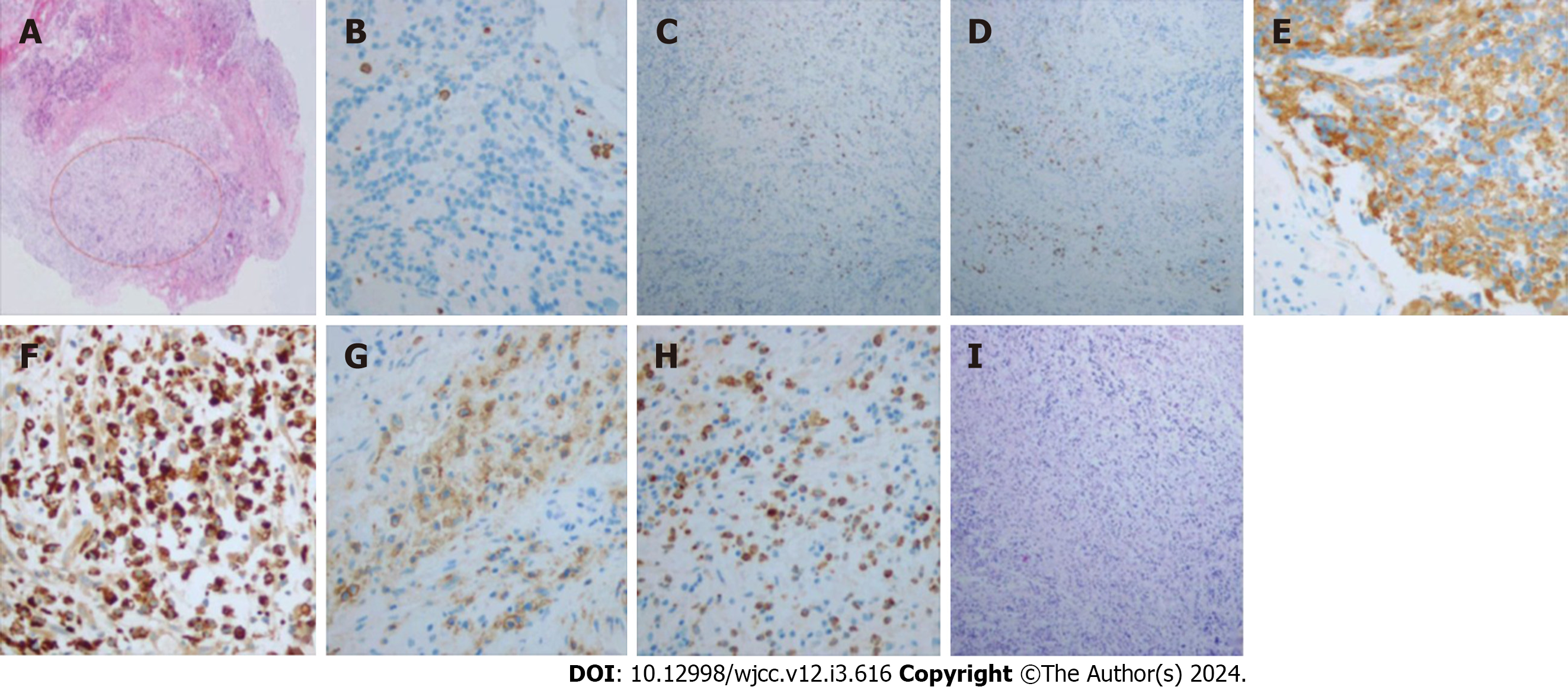
Figure 2 Histopathological analysis and immunohistochemical examination of the extracted specimen.
A: Hematoxylin and eosin staining (× 40); B: Immunohistochemical staining for showing CD3 positive staining (× 200); C: CD4 positive staining (× 100); D: CD20 positive staining (× 100); E: CD56 positive staining (× 400); F: CD68 positive staining (× 400); G: CD138 positive staining (× 400); H: MPO positive staining (× 400); I: PAS negative staining (× 100).
- Citation: Zhu XM, Dong CX, Xie L, Liu HX, Hu HQ. Brain abscess from oral microbiota approached by metagenomic next-generation sequencing: A case report and review of literature. World J Clin Cases 2024; 12(3): 616-622
- URL: https://www.wjgnet.com/2307-8960/full/v12/i3/616.htm
- DOI: https://dx.doi.org/10.12998/wjcc.v12.i3.616


