
















©The Author(s) 2024.
World J Clin Cases. Aug 16, 2024; 12(23): 5431-5440
Published online Aug 16, 2024. doi: 10.12998/wjcc.v12.i23.5431
Published online Aug 16, 2024. doi: 10.12998/wjcc.v12.i23.5431
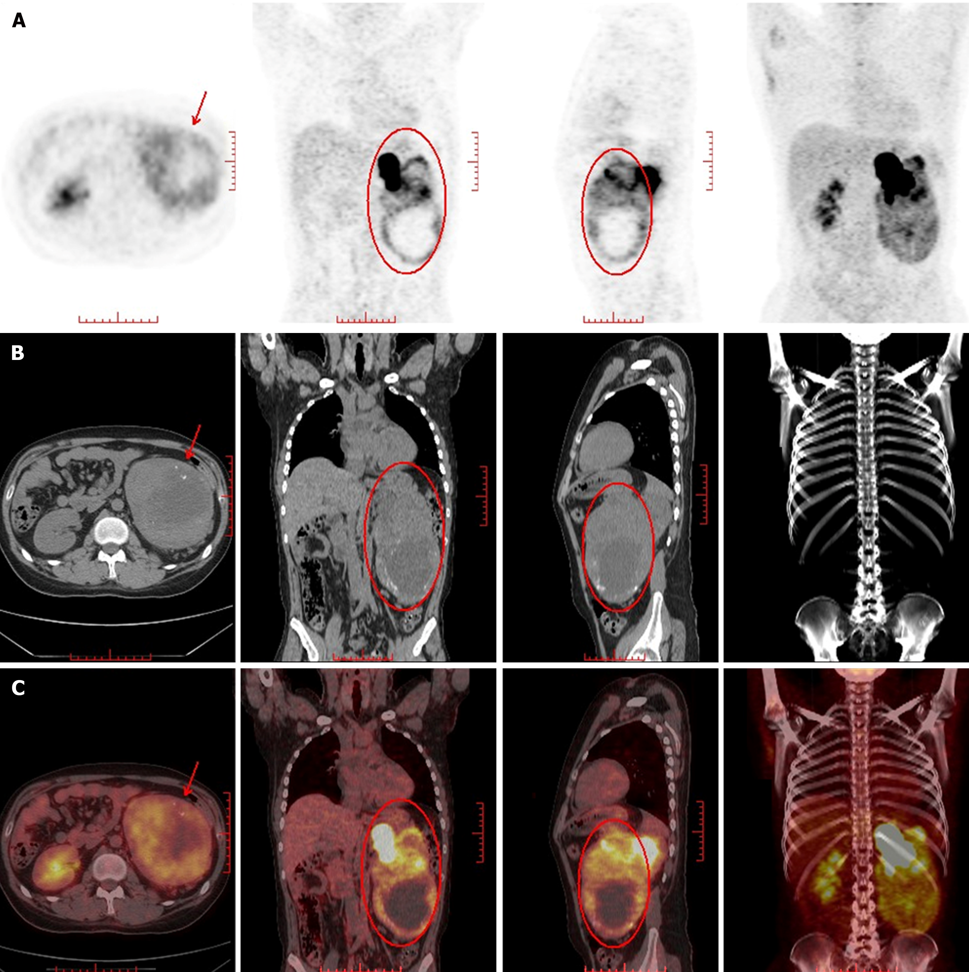
Figure 3 Nuclear medicine image of the primary tumor.
A: Increased radioactivity in the left kidney; B: A large mass of mixed density was found in the left kidney. The large cross section of the lesion was about 11.8 cm × 9.7 cm; C: The solid component showed uneven radioactive concentration, Maximum Standardized Uptake Value 10.4, the left side of the kidney was compressed and displaced upward, and the renal calyx was dilated and hydronephrosis.
- Citation: Dong GF, Hou YK, Ma Q, Ma SY, Wang YJ, Rexiati M, Wang WG. Cushing's syndrome caused by giant Ewing's sarcoma of the kidney: A case report and review of literature. World J Clin Cases 2024; 12(23): 5431-5440
- URL: https://www.wjgnet.com/2307-8960/full/v12/i23/5431.htm
- DOI: https://dx.doi.org/10.12998/wjcc.v12.i23.5431