
















©The Author(s) 2024.
World J Clin Cases. Jul 26, 2024; 12(21): 4813-4819
Published online Jul 26, 2024. doi: 10.12998/wjcc.v12.i21.4813
Published online Jul 26, 2024. doi: 10.12998/wjcc.v12.i21.4813
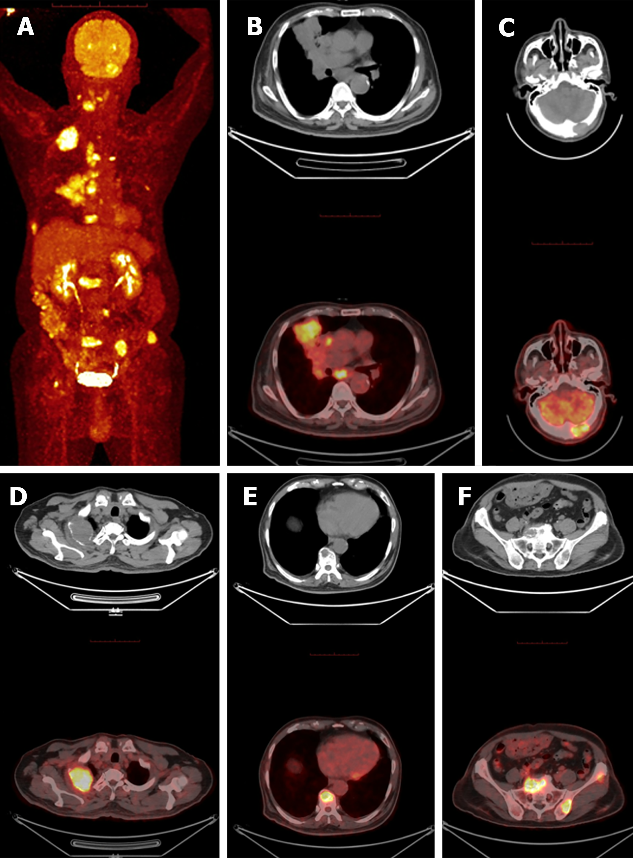
Figure 3 Positron emission tomography/computed tomography.
A-F: Positron emission tomography/computed tomography showed huge hyper
- Citation: Mo YJ, Lin LN, Tao JL, Zhang T, Zhang JH. Hepatoid adenocarcinoma of the lung: A case report. World J Clin Cases 2024; 12(21): 4813-4819
- URL: https://www.wjgnet.com/2307-8960/full/v12/i21/4813.htm
- DOI: https://dx.doi.org/10.12998/wjcc.v12.i21.4813