
















©The Author(s) 2024.
World J Clin Cases. Jul 26, 2024; 12(21): 4777-4782
Published online Jul 26, 2024. doi: 10.12998/wjcc.v12.i21.4777
Published online Jul 26, 2024. doi: 10.12998/wjcc.v12.i21.4777
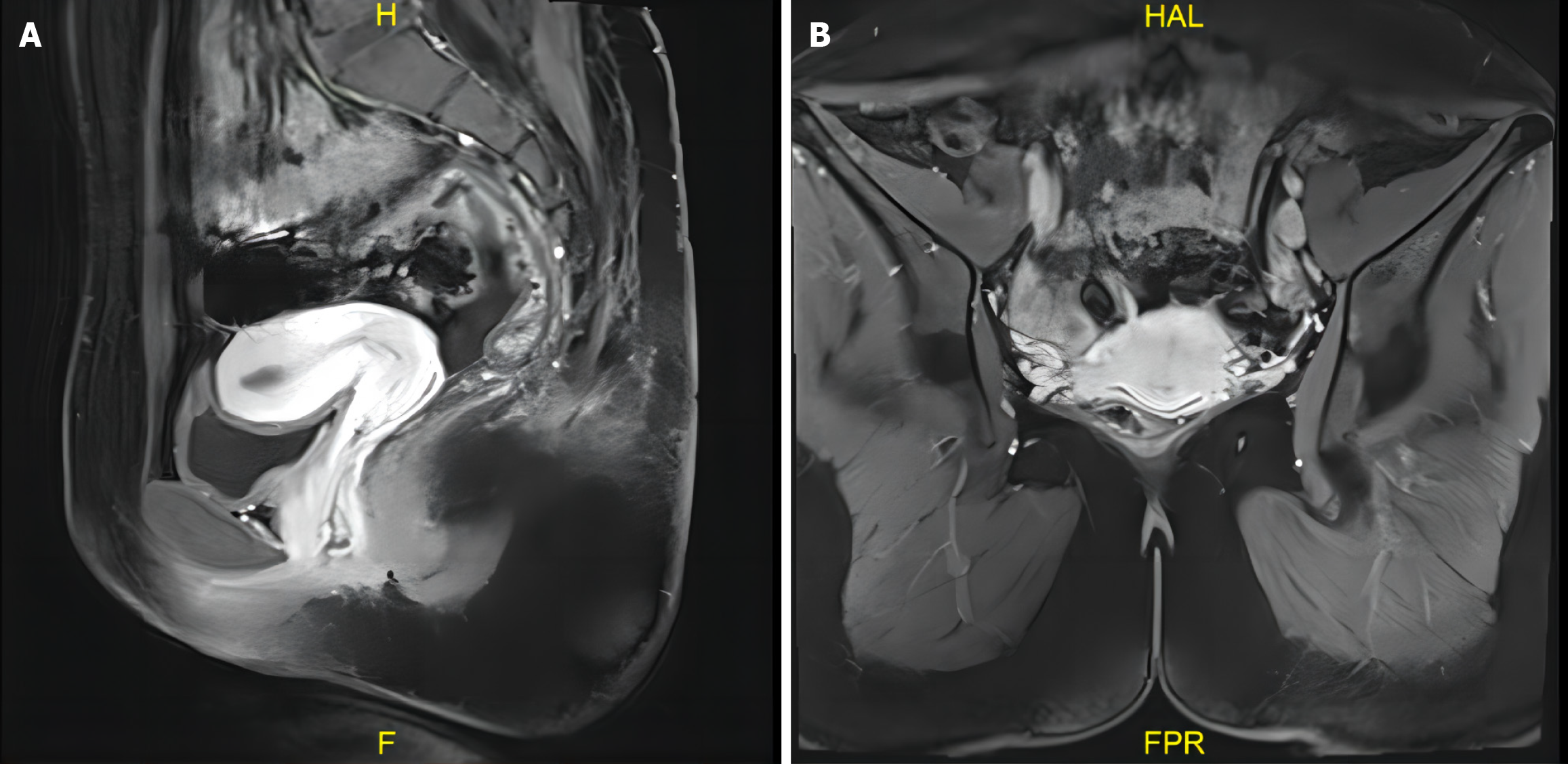
Figure 3 Pelvic magnetic resonance imaging revealed a normal uterus and cervix.
A and B: Sagittal (A) and axial (B) pelvic magnetic resonance imaging showed normal uterine size and shape, no abnormal signals, and no obvious abnormal signal shadows in the bilateral adnexa.
- Citation: Li H, Mei SS, Mao PY, Wang XY, Yang HD. Hysteroscopic cervical biopsy for women with persistent human papillomavirus infection after loop electrosurgical excision procedure: A case report. World J Clin Cases 2024; 12(21): 4777-4782
- URL: https://www.wjgnet.com/2307-8960/full/v12/i21/4777.htm
- DOI: https://dx.doi.org/10.12998/wjcc.v12.i21.4777