Copyright
©The Author(s) 2024.
World J Clin Cases. Jan 16, 2024; 12(2): 451-459
Published online Jan 16, 2024. doi: 10.12998/wjcc.v12.i2.451
Published online Jan 16, 2024. doi: 10.12998/wjcc.v12.i2.451
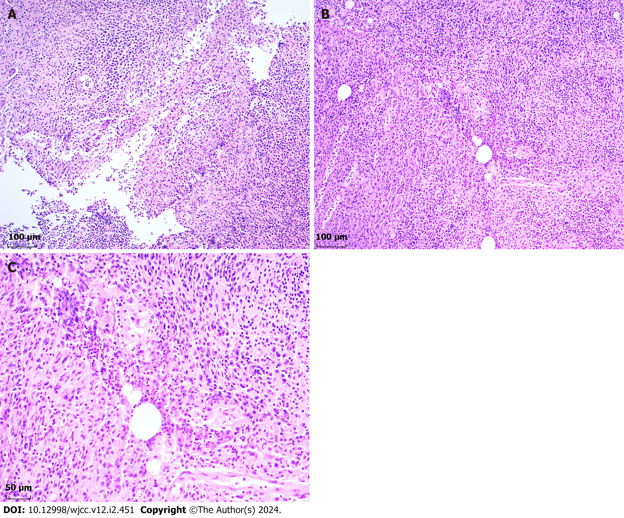
Figure 3 Pathological investigation revealed a poorly formed non-caseating granuloma.
A: Depicting the poorly formed non-caseating granuloma encircled by epithelioid cells, lymphocytes, plasma cells, neutrophils, and multinucleate giant cells. [hematoxylin and eosin (HE), 100 × magnification]; B: Illustrating the non-caseating granuloma centered on a vacuolated space (HE, 100 × magnification); C: High-resolution view of Figure 3B, highlighting the vacuolated space rimmed by neutrophils and surrounded by epithelioid cells, lymphocytes, plasma cells, and multinucleate giant cells (HE, 200 × magnification).
- Citation: Cui LY, Sun CP, Li YY, Liu S. Granulomatous mastitis in a 50-year-old male: A case report and review of literature. World J Clin Cases 2024; 12(2): 451-459
- URL: https://www.wjgnet.com/2307-8960/full/v12/i2/451.htm
- DOI: https://dx.doi.org/10.12998/wjcc.v12.i2.451


