Copyright
©The Author(s) 2024.
World J Clin Cases. Jan 16, 2024; 12(2): 354-360
Published online Jan 16, 2024. doi: 10.12998/wjcc.v12.i2.354
Published online Jan 16, 2024. doi: 10.12998/wjcc.v12.i2.354
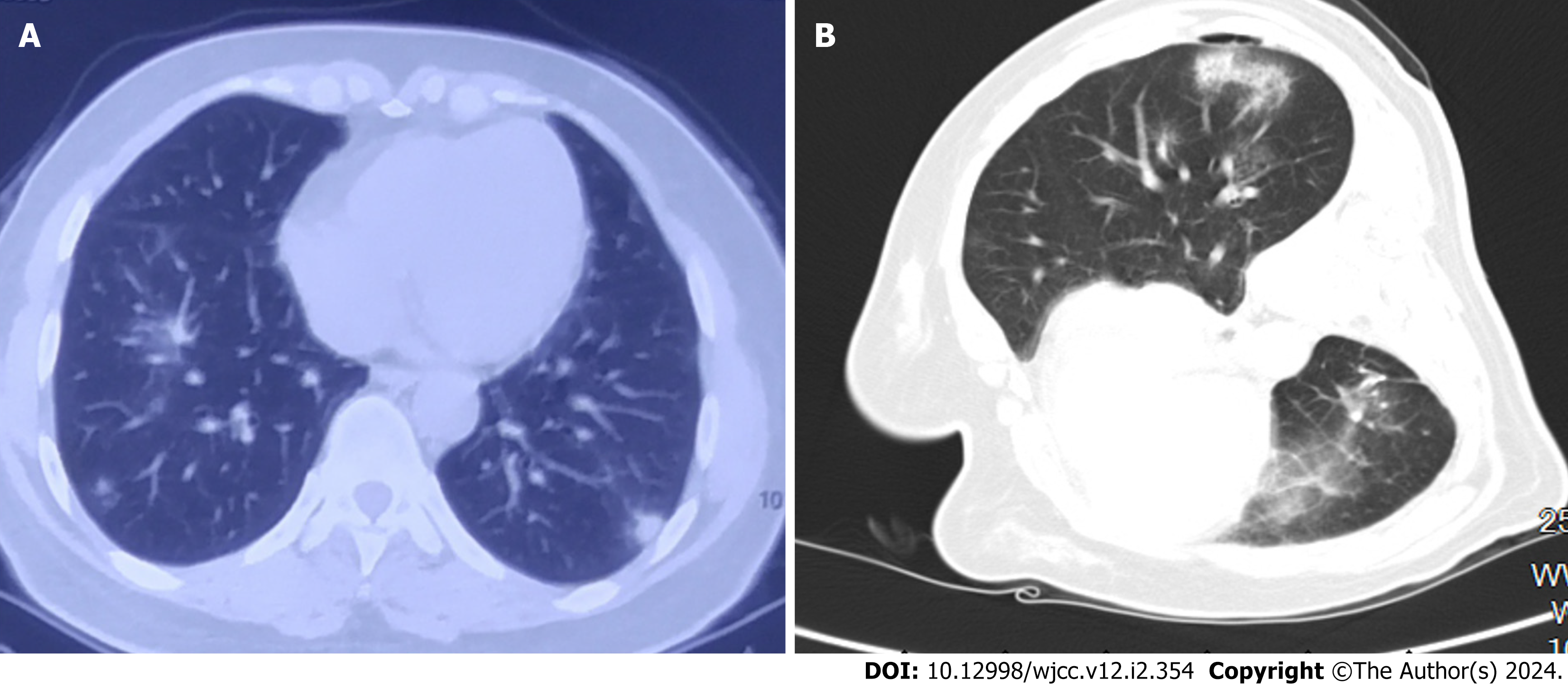
Figure 1 Lung imaging before and after lobectomy.
A: Chest computed tomography (CT) showed two high-density pulmonary nodules in the posterior basal segment of the left lower lobe; B: Chest CT revealed a new pulmonary nodule in the right lower lobe (left lateral position).
- Citation: Feng SL, Li JY, Dong CL. Primary biliary cholangitis presenting with granulomatous lung disease misdiagnosed as lung cancer: A case report. World J Clin Cases 2024; 12(2): 354-360
- URL: https://www.wjgnet.com/2307-8960/full/v12/i2/354.htm
- DOI: https://dx.doi.org/10.12998/wjcc.v12.i2.354


