
















©The Author(s) 2024.
World J Clin Cases. Jun 16, 2024; 12(17): 3130-3137
Published online Jun 16, 2024. doi: 10.12998/wjcc.v12.i17.3130
Published online Jun 16, 2024. doi: 10.12998/wjcc.v12.i17.3130
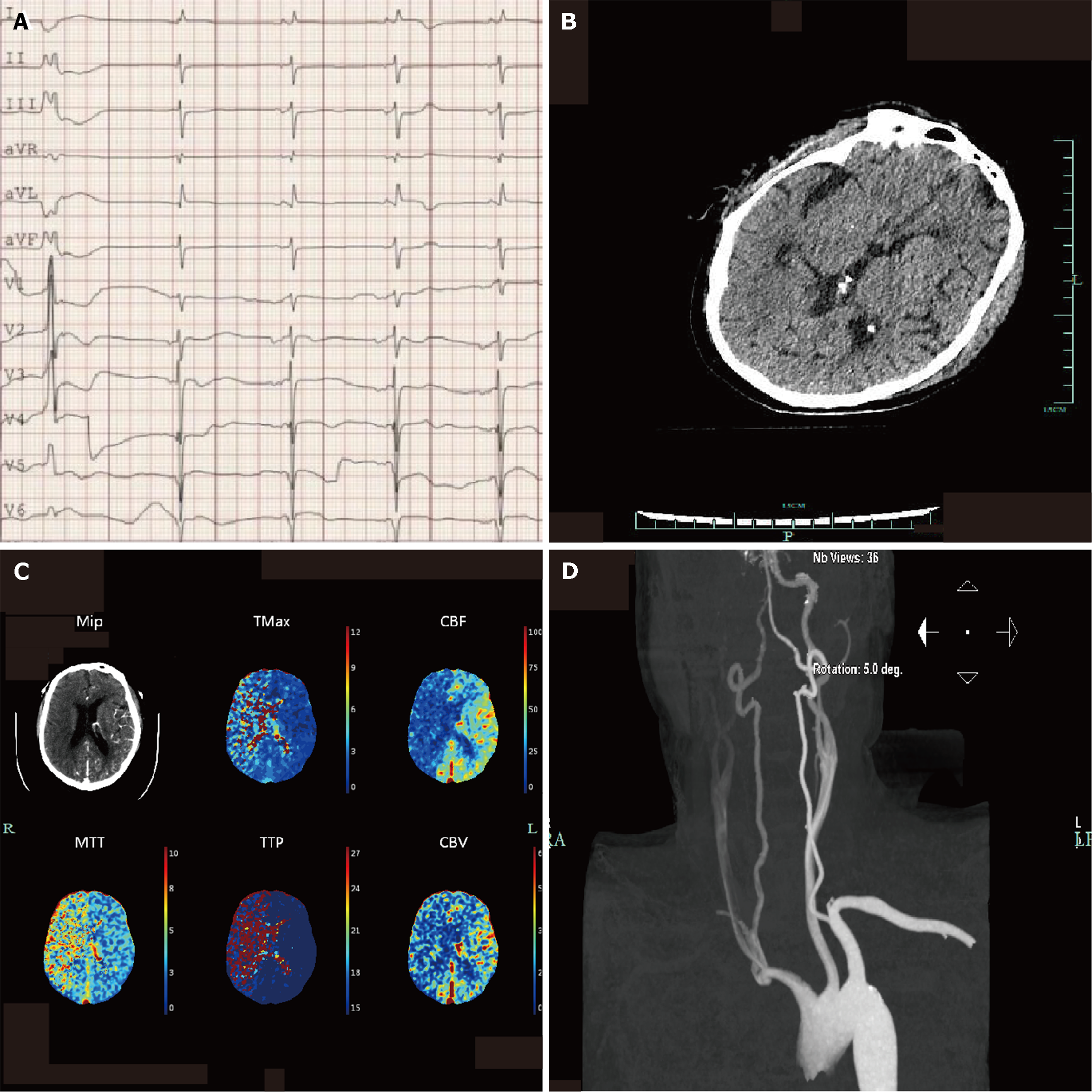
Figure 1 The initial neurological assessment on the day of admission could not confirm the patient's mental state.
After the administration of naloxone and flumazenil, the Glasgow Coma Scale score was 2 + endotracheal tube + 3, and we completed further imaging examination. A: Electrocardiogram showing sinus rhythm, atrial rhythm, premature ventricular beats, occasional junctional rhythm, acute anterior wall and anterior wall myocardial infarction, and atrioventricular block; B: Head computed tomography images showing no obvious signs of stroke; C: Computed tomography perfusion indicating cerebral infarction and poor compensatory blood flow and ischemic hypoperfusion in the right large frontoparietal and temporal lobe; D: Carotid artery computed tomography angiography image showing sparse blood flow in the right common carotid artery, M1 segment of the right middle cerebral artery, and vertebral artery blood.
- Citation: Xu M, Yan JY, Jin JJ, Li T. Cerebral pseudoinfarction due to venoarterial extracorporeal membrane oxygenation: A case report. World J Clin Cases 2024; 12(17): 3130-3137
- URL: https://www.wjgnet.com/2307-8960/full/v12/i17/3130.htm
- DOI: https://dx.doi.org/10.12998/wjcc.v12.i17.3130