
















©The Author(s) 2024.
World J Clin Cases. May 26, 2024; 12(15): 2678-2681
Published online May 26, 2024. doi: 10.12998/wjcc.v12.i15.2678
Published online May 26, 2024. doi: 10.12998/wjcc.v12.i15.2678
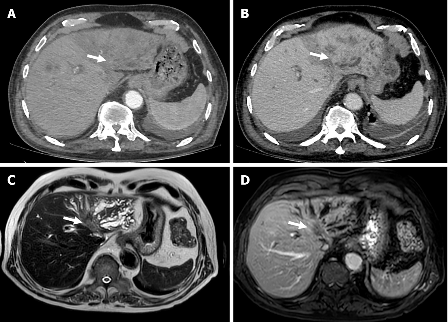
Figure 2 69-years-old woman with biliary obstruction secondary to intrahepatic mass-forming type cholangiocarcinoma with tumoral involvement of the biliary ducts confluence.
A: Axial late computed tomography arterial phase depicts a soft-tissue mass in left hepatic lobe, with delayed phase enhancement; B: Dilatation of intrahepatic ducts; C: Axial magnetic resonance images demonstrate a T2-weighted images slightly hyperintense; D: T1-weighted images hypointense heterogeneous mass occluding the confluence of the hepatic ducts with moderate dilatation of left lobar intrahepatic bile ducts, and tumoral involvement of the left intrahepatic biliary branches.
- Citation: Lindner C. Imaging features of malignant vs stone-induced biliary obstruction: Aspects to consider. World J Clin Cases 2024; 12(15): 2678-2681
- URL: https://www.wjgnet.com/2307-8960/full/v12/i15/2678.htm
- DOI: https://dx.doi.org/10.12998/wjcc.v12.i15.2678