Copyright
©The Author(s) 2024.
World J Clin Cases. Apr 26, 2024; 12(12): 2065-2073
Published online Apr 26, 2024. doi: 10.12998/wjcc.v12.i12.2065
Published online Apr 26, 2024. doi: 10.12998/wjcc.v12.i12.2065
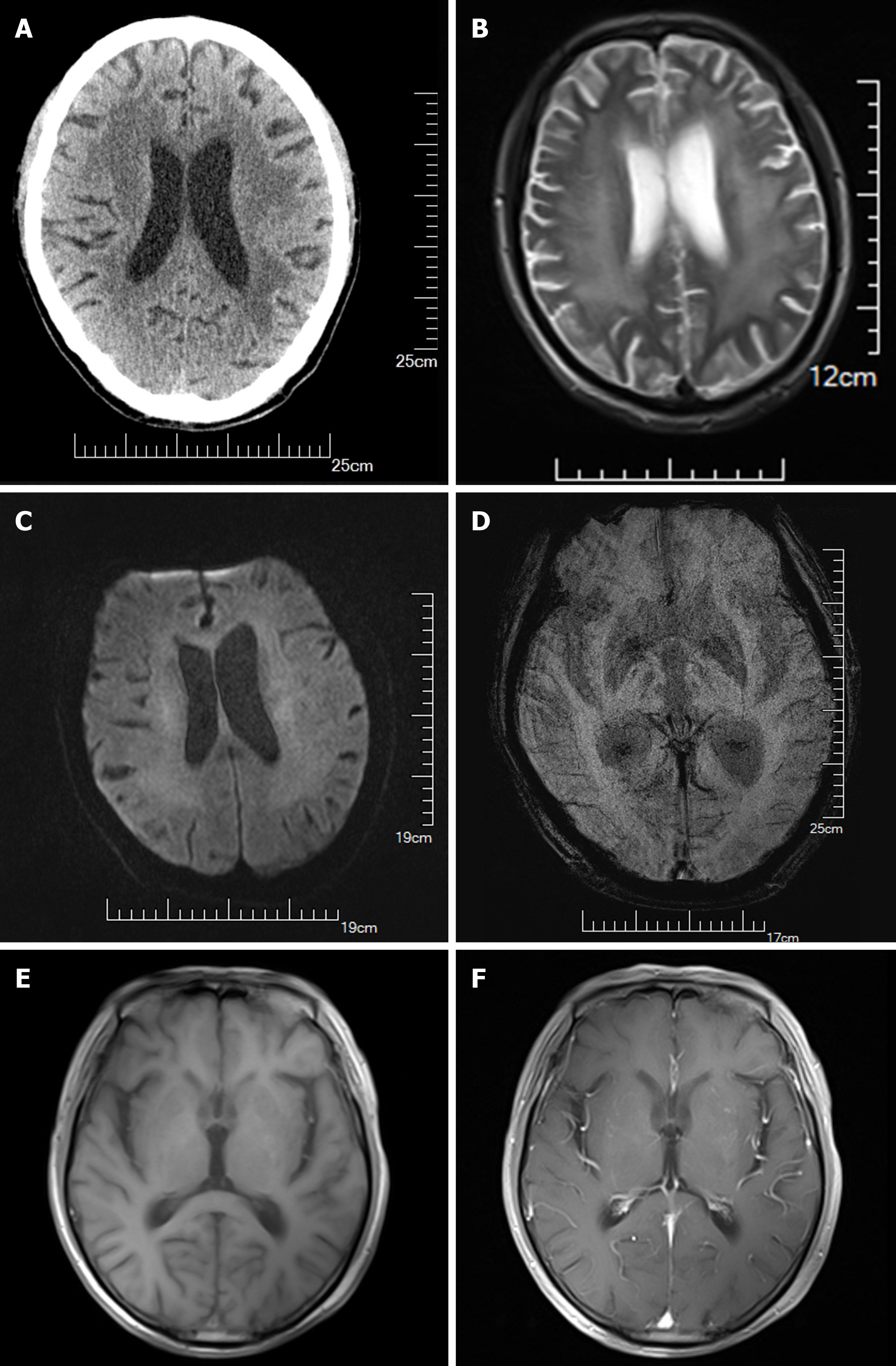
Figure 2 Pre- and post-treatment cranial imaging examinations of the patient.
A-D: Neuroradiological presentation of this patient before treatment; The brain computed tomography reveals encephalatrophy and demyelination in the white matter (A); Axial T1-weighted brain magnetic resonance imaging (MRI) with pre-contrast shows mild encephalatrophy and demyelination around the bilateral cerebral ventricles (B); Axial T1-weighted brain MRI with post-contrast displays no enhancement of any lesions (C and D); E and F: Neuroradiological presentation of this patient after treatment; Axial T1-weighted brain MRI with pre-contrast illustrates slight encephalatrophy and the significantly improved demyelination in the white matter around the bilateral cerebral ventricles (E); Axial T1-weighted brain MRI with post-contrast demonstrates no enhancement in any of the lesions (F).
- Citation: He YS, Qin XH, Feng M, Huang QJ, Zhang MJ, Guo LL, Bao MB, Tao Y, Dai HY, Wu B. Human immunodeficiency virus-associated dementia complex with positive 14-3-3 protein in cerebrospinal fluid: A case report. World J Clin Cases 2024; 12(12): 2065-2073
- URL: https://www.wjgnet.com/2307-8960/full/v12/i12/2065.htm
- DOI: https://dx.doi.org/10.12998/wjcc.v12.i12.2065


