Copyright
©The Author(s) 2024.
World J Clin Cases. Jan 6, 2024; 12(1): 210-216
Published online Jan 6, 2024. doi: 10.12998/wjcc.v12.i1.210
Published online Jan 6, 2024. doi: 10.12998/wjcc.v12.i1.210
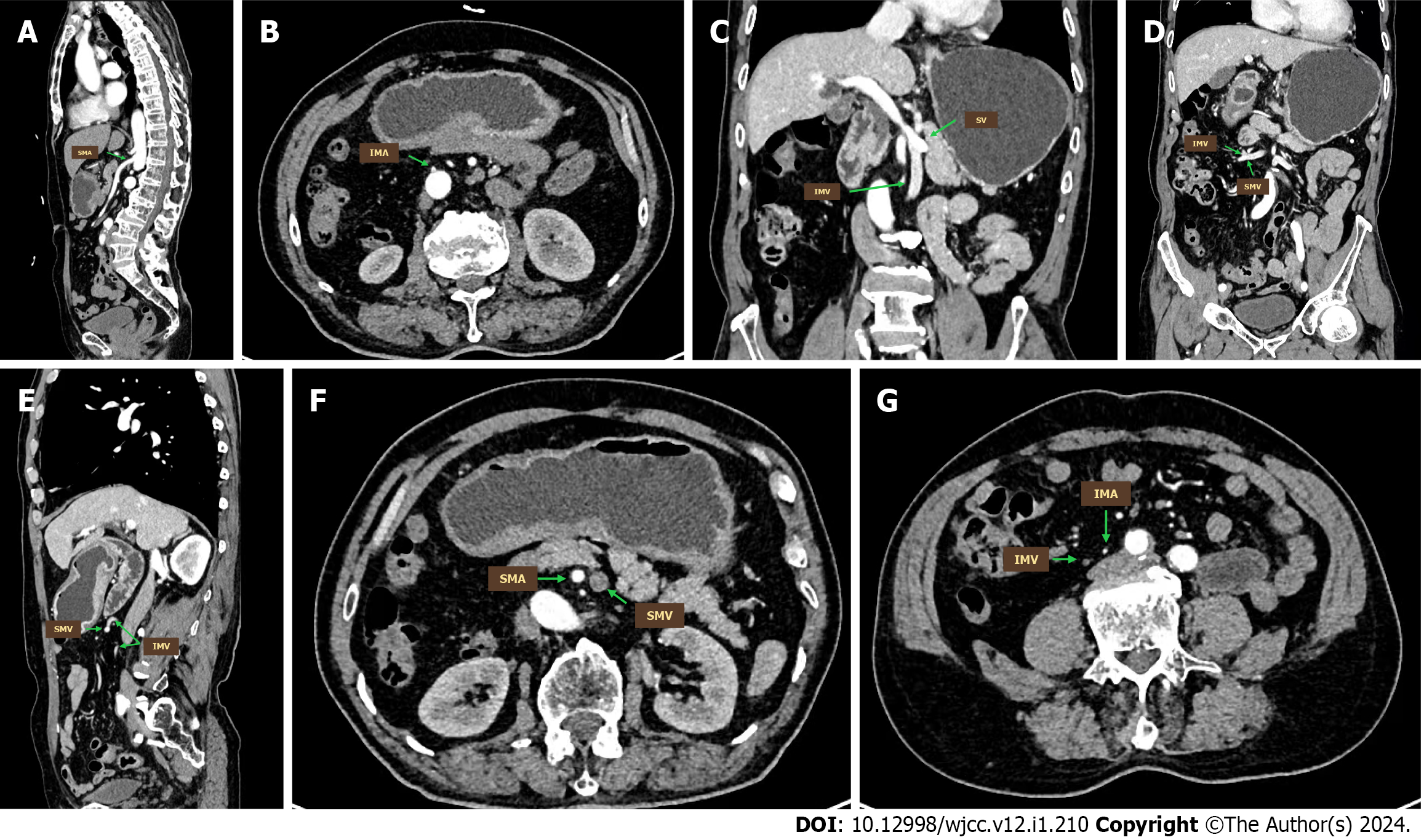
Figure 2 Reconstruction images of the abdomen.
A: Sagittal reconstruction of the computed tomography (CT) images showed the superior mesenteric artery (SMA) emanated from the abdominal aorta; B: Transverse reconstruction of the CT images showed the inferior mesenteric artery (IMA) originating from the abdominal aorta; C: Coronal reconstruction of the CT images showed the splenic vein joining the superior mesenteric vein (SMV), forming the portal vein; D: Coronal reconstruction of the CT images showed the inferior mesenteric vein (IMV) joining the SMV on the right side of the abdominal aorta; E: Coronal reconstruction of the CT images showed the IMV converging into the SMV as a result of mesentery malrotation; F: Transverse reconstruction of the CT images showed the SMA on the right to the SMV; G: Transverse reconstruction of the CT images showed the IMA and IMV running alongside the right side of the common iliac artery. SMA: Superior mesenteric artery; IMA: Inferior mesenteric artery; SV: Splenic vein; SMV: Superior mesenteric vein; IMV: Inferior mesenteric vein.
- Citation: Jia XH, Kong S, Gao XX, Cong BC, Zheng CN. Intestinal malrotation complicated with gastric cancer: A case report. World J Clin Cases 2024; 12(1): 210-216
- URL: https://www.wjgnet.com/2307-8960/full/v12/i1/210.htm
- DOI: https://dx.doi.org/10.12998/wjcc.v12.i1.210


