Copyright
©The Author(s) 2023.
World J Clin Cases. Mar 26, 2023; 11(9): 2074-2083
Published online Mar 26, 2023. doi: 10.12998/wjcc.v11.i9.2074
Published online Mar 26, 2023. doi: 10.12998/wjcc.v11.i9.2074
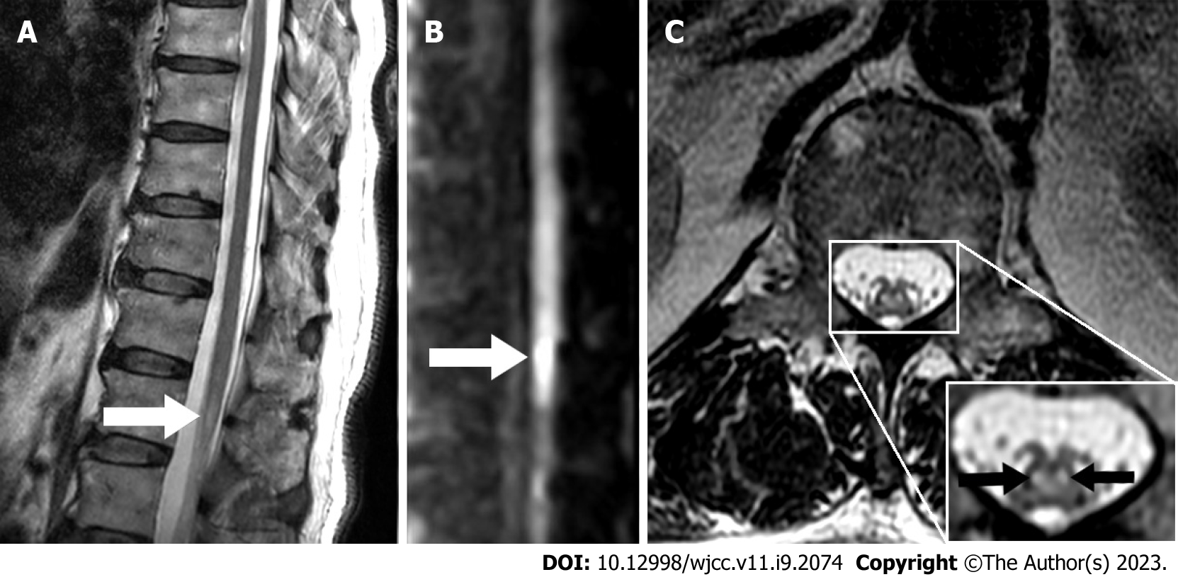
Figure 1 Conus medullaris magnetic resonance exam and “snake-eye appearance”.
A: The arrow shows conus infarction on T2 weighted magnetic resonance imaging (MRI) (sagittal position); B: The arrow shows conus infarction on diffusion-weighted imaging image (sagittal position); C: The arrows show “snake-eye appearance” on T2 weighted MRI (axial position).
- Citation: Zhang QY, Xu LY, Wang ML, Cao H, Ji XF. Spontaneous conus infarction with "snake-eye appearance" on magnetic resonance imaging: A case report and literature review. World J Clin Cases 2023; 11(9): 2074-2083
- URL: https://www.wjgnet.com/2307-8960/full/v11/i9/2074.htm
- DOI: https://dx.doi.org/10.12998/wjcc.v11.i9.2074


