Copyright
©The Author(s) 2023.
World J Clin Cases. Mar 16, 2023; 11(8): 1847-1856
Published online Mar 16, 2023. doi: 10.12998/wjcc.v11.i8.1847
Published online Mar 16, 2023. doi: 10.12998/wjcc.v11.i8.1847
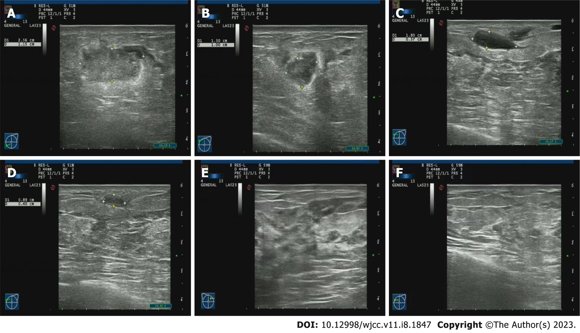
Figure 1 Imaging examinations.
A: August 30, 2022: Several hypoechoic nodules can be seen in the left breast, the larger ones are about 2.6 cm × 1.2 cm (in the direction of 8 points) and 1.3 cm × 1.0 cm (in the direction of 10 points); B: August 30, 2022: Several cystic nodules can be seen in the right breast, the larger size is about 0.4 cm × 0.4 cm (in the direction of 10 points), and the internal sound transmission is poor; C: September 5, 2022: Several hypoechoic masses can be seen in the left breast, the larger size is about 1.8 cm × 0.6 cm (in the direction of 10 points); D: September 12, 2022: Several hypoechoic masses can be seen in the left breast, the larger one is about 0.9 cm × 0.4 cm (in the direction of 10 points); E and F: September 27, 2022: Bilateral mammary glands have normal morphology.
- Citation: Jin LH, Zheng HL, Lin YX, Yang Y, Liu JL, Li RL, Ye HJ. Lactation breast abscess treated with Gualou Xiaoyong decoction and painless lactation manipulation: A case report and review of literature. World J Clin Cases 2023; 11(8): 1847-1856
- URL: https://www.wjgnet.com/2307-8960/full/v11/i8/1847.htm
- DOI: https://dx.doi.org/10.12998/wjcc.v11.i8.1847


