Copyright
©The Author(s) 2023.
World J Clin Cases. Mar 16, 2023; 11(8): 1808-1813
Published online Mar 16, 2023. doi: 10.12998/wjcc.v11.i8.1808
Published online Mar 16, 2023. doi: 10.12998/wjcc.v11.i8.1808
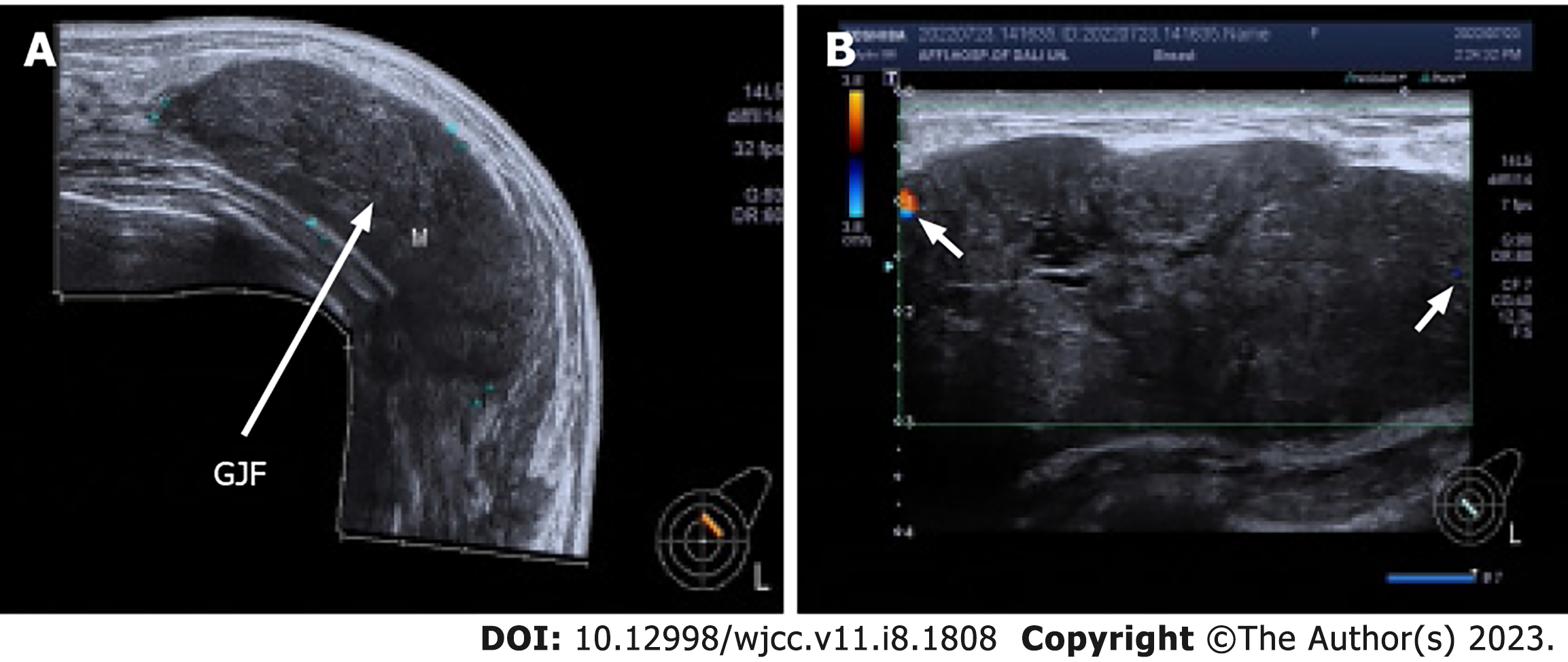
Figure 1 Breast ultrasonography.
A: Panoramic ultrasound imaging reveals the left breast with a large benign mass (long arrow); B: Punctate Doppler flow signals (short arrow) at the edge of the breast mass.
- Citation: Wang J, Zhang DD, Cheng JM, Chen HY, Yang RJ. Giant juvenile fibroadenoma in a 14-year old Chinese female: A case report. World J Clin Cases 2023; 11(8): 1808-1813
- URL: https://www.wjgnet.com/2307-8960/full/v11/i8/1808.htm
- DOI: https://dx.doi.org/10.12998/wjcc.v11.i8.1808


