Copyright
©The Author(s) 2023.
World J Clin Cases. Mar 6, 2023; 11(7): 1560-1568
Published online Mar 6, 2023. doi: 10.12998/wjcc.v11.i7.1560
Published online Mar 6, 2023. doi: 10.12998/wjcc.v11.i7.1560
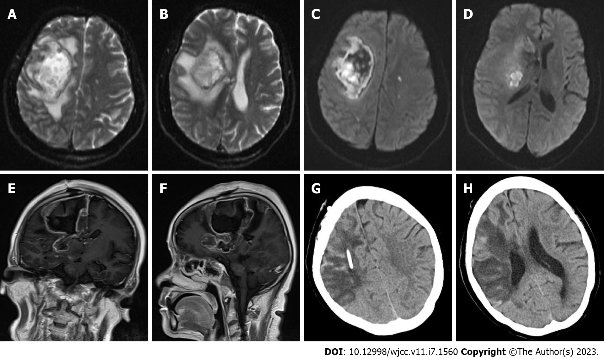
Figure 3 Head magnetic resonance imaging and postoperative head computed tomography review.
A-D: Flaky T2 mixed signal was observed in the right frontal lobe and basal ganglia, there was a mixed signal on diffusion-weighted imaging (DWI), and the right lateral ventricle was compressed. In the left cerebral hemisphere, multiple long T2 signals were observed, some of which were high on DWI, and the midline was shifted to the left; E and F: After enhancement, partial ring enhancement was observed; G and H: On the 10th d after the brain lesion resection, repeat head computed tomography showed a large low-density shadow, and the midline returned to normal.
- Citation: Chen CH, Chen JN, Du HG, Guo DL. Isolated cerebral mucormycosis that looks like stroke and brain abscess: A case report and review of the literature. World J Clin Cases 2023; 11(7): 1560-1568
- URL: https://www.wjgnet.com/2307-8960/full/v11/i7/1560.htm
- DOI: https://dx.doi.org/10.12998/wjcc.v11.i7.1560


