Copyright
©The Author(s) 2023.
World J Clin Cases. Mar 6, 2023; 11(7): 1528-1548
Published online Mar 6, 2023. doi: 10.12998/wjcc.v11.i7.1528
Published online Mar 6, 2023. doi: 10.12998/wjcc.v11.i7.1528
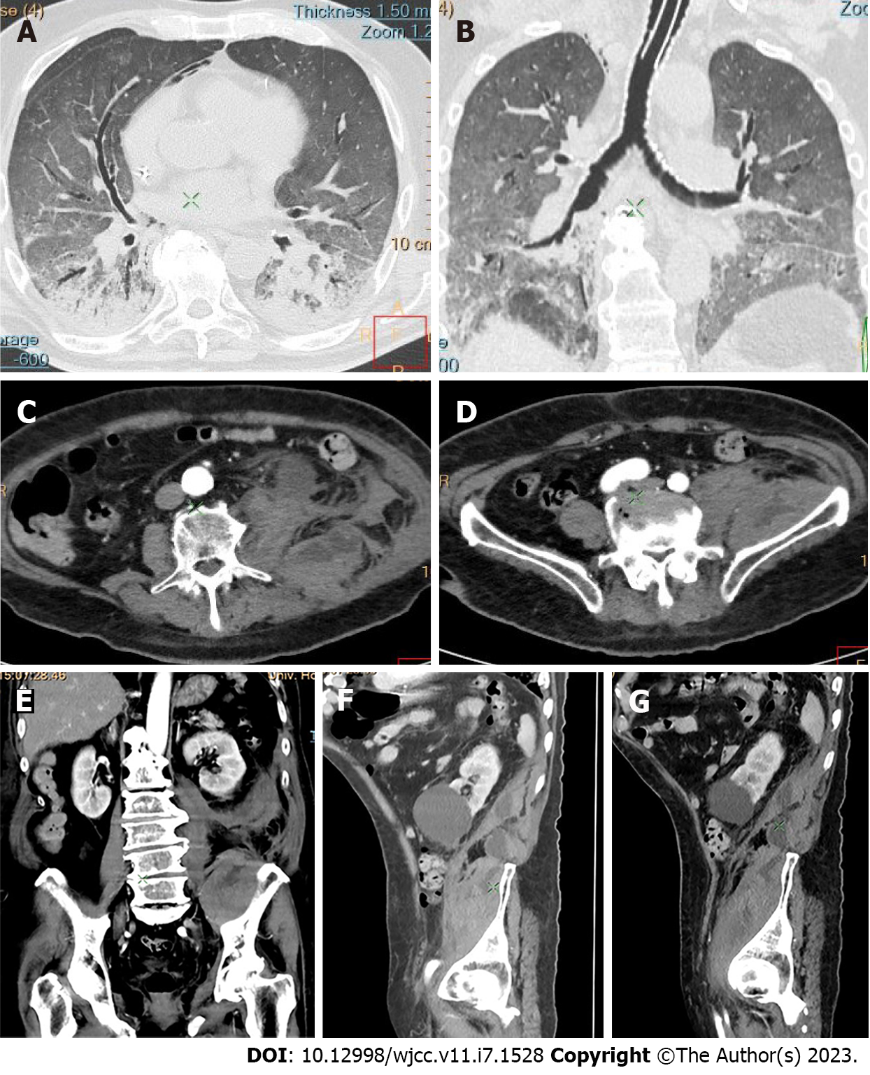
Figure 5 Abdominal and pelvic computed tomography and dynamic contrast enhanced computed tomography of retroperitoneal hemorrhage on the left.
A: Bilateral lung consolidations with posterior predilection, predominantly involving the lower lobes, including “ground glass” opacities and vascular enlargement in cross section; B: Bilateral lung consolidations with posterior predilection, predominantly involving the lower lobes, including “ground glass” opacities and vascular enlargement in vertical section; C-E: Retroperitoneal hemorrhagic collection in the left flank and iliac fossa by postcontrast computed tomography axial and coronal images. Axial and coronal reformat images of dilatation of the psoas muscles; F and G: Sagittal reconstruction plane of the entire length of the iliac muscle affected by a hematoma.
- Citation: Evrev D, Sekulovski M, Gulinac M, Dobrev H, Velikova T, Hadjidekov G. Retroperitoneal and abdominal bleeding in anticoagulated COVID-19 hospitalized patients: Case series and brief literature review. World J Clin Cases 2023; 11(7): 1528-1548
- URL: https://www.wjgnet.com/2307-8960/full/v11/i7/1528.htm
- DOI: https://dx.doi.org/10.12998/wjcc.v11.i7.1528


