Copyright
©The Author(s) 2023.
World J Clin Cases. Mar 6, 2023; 11(7): 1528-1548
Published online Mar 6, 2023. doi: 10.12998/wjcc.v11.i7.1528
Published online Mar 6, 2023. doi: 10.12998/wjcc.v11.i7.1528
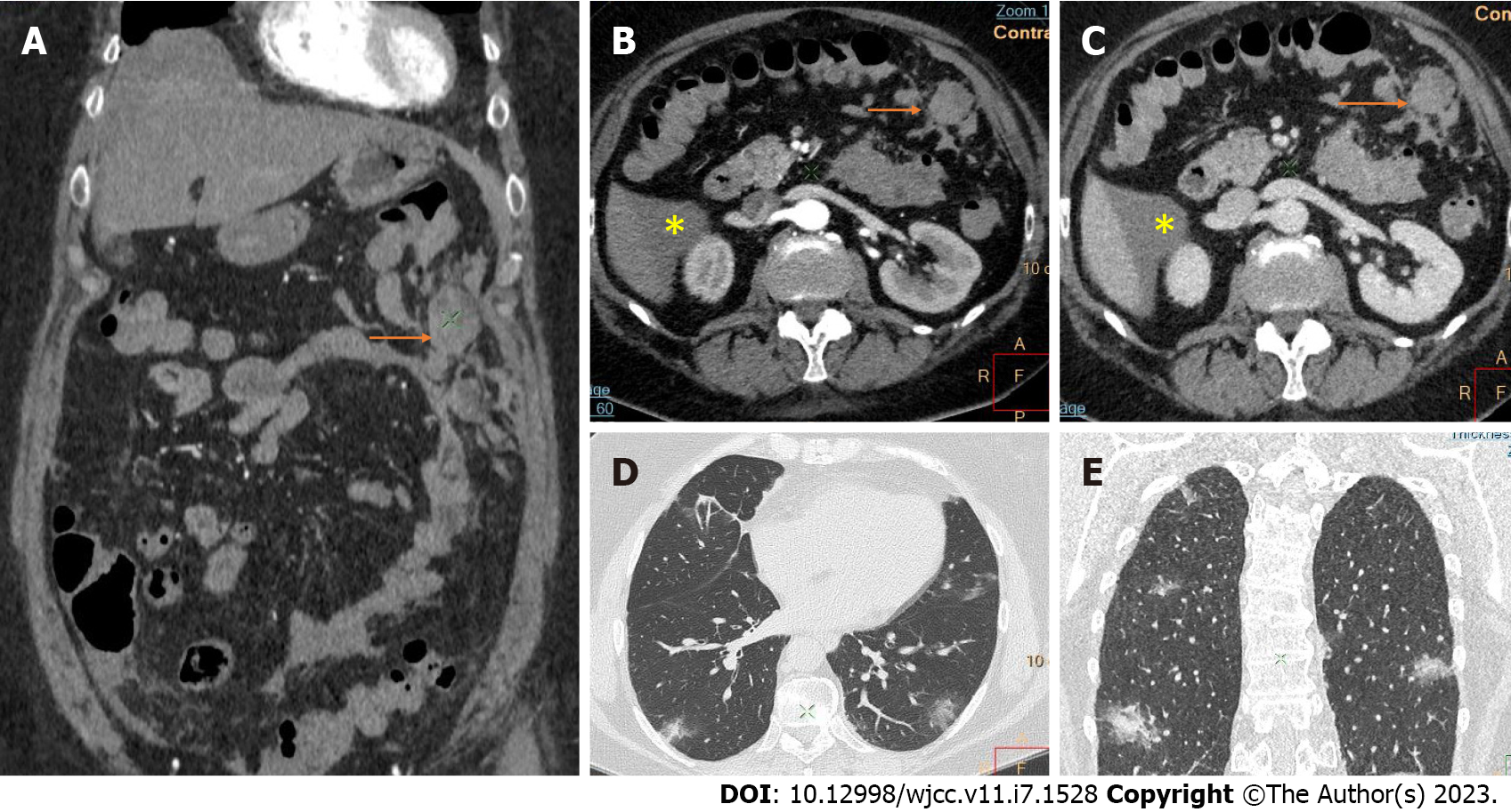
Figure 2 Computed tomography images of intraabdominal hemorrhage and lung involvement by coronavirus disease 2019 pneumonia.
A: Coronal reconstruction; B and C: Axial images with contrast demonstrate an intraabdominal hemorrhagic collection in the left upper quadrant without clear visualization of the site of bleeding (arrows) and small ascites close to the lower pole of the liver (asterisk); D and E: Bilateral lung "ground glass" consolidations in coronavirus disease 2019 pneumonia.
- Citation: Evrev D, Sekulovski M, Gulinac M, Dobrev H, Velikova T, Hadjidekov G. Retroperitoneal and abdominal bleeding in anticoagulated COVID-19 hospitalized patients: Case series and brief literature review. World J Clin Cases 2023; 11(7): 1528-1548
- URL: https://www.wjgnet.com/2307-8960/full/v11/i7/1528.htm
- DOI: https://dx.doi.org/10.12998/wjcc.v11.i7.1528


